González-Fernández Bárbara, Sánchez Diana I, Crespo Irene, San-Miguel Beatriz, de Urbina Juan Ortiz, González-Gallego Javier, Tuñón María J
Institute of Biomedicine (IBIOMED), University of León, León, Spain.
Centro de Investigación Biomédica en Red de Enfermedades Hepáticas y Digestivas (CIBERehd), León, Spain.
Front Pharmacol. 2018 May 28;9:556. doi: 10.3389/fphar.2018.00556. eCollection 2018.
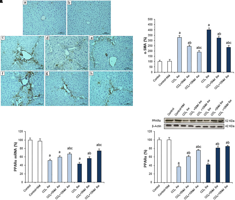
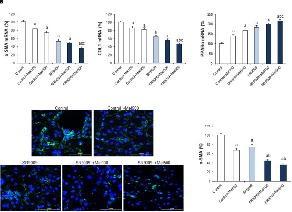
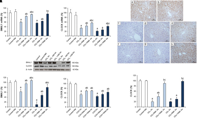
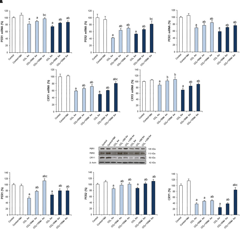
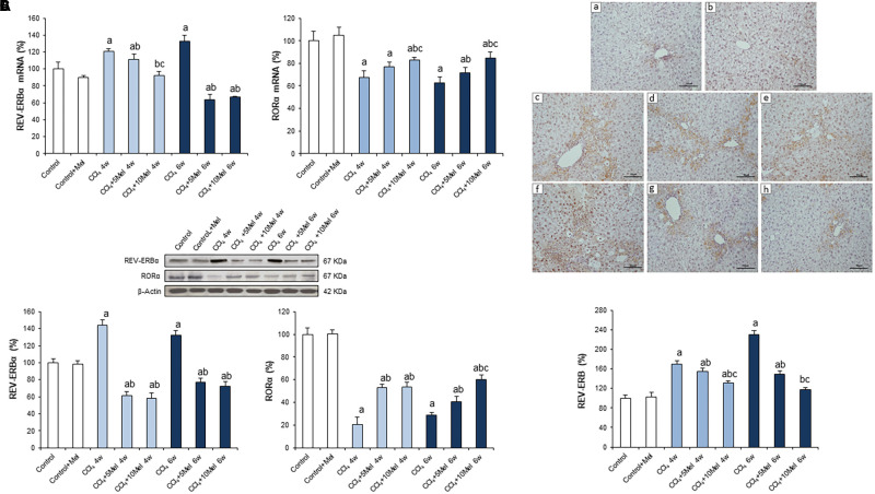
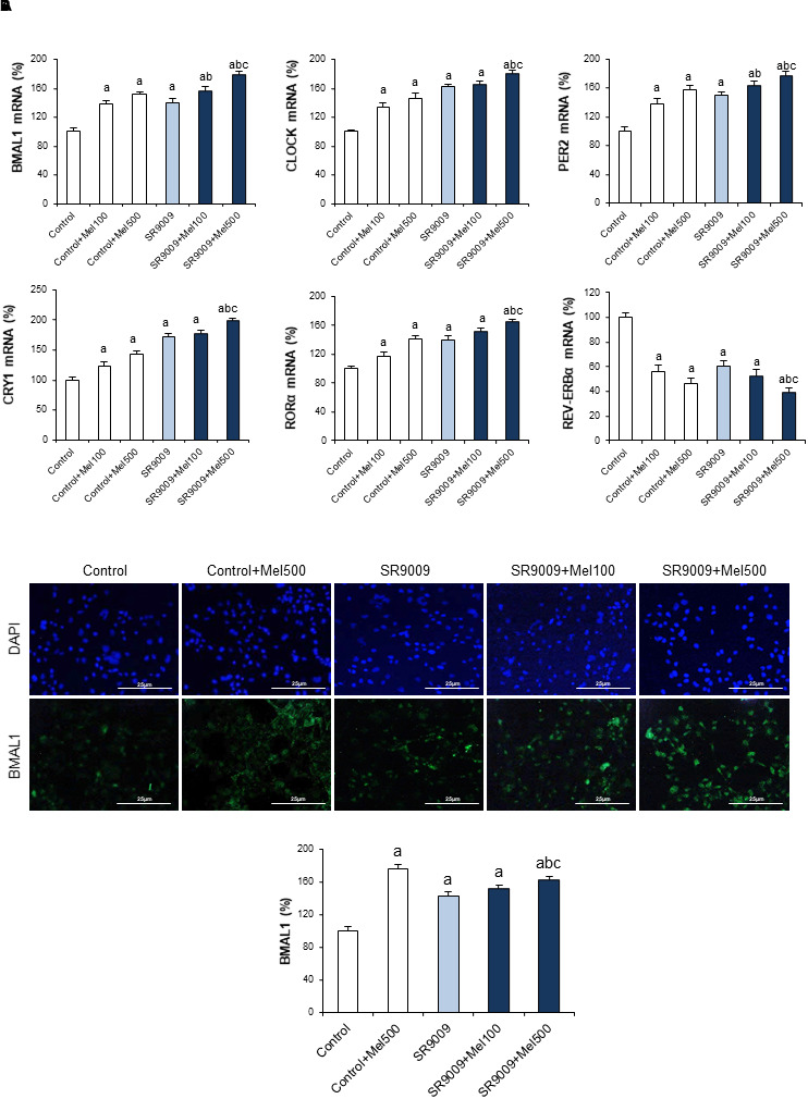
Dysregulation of the circadian clock machinery is a critical mechanism in the pathogenesis of fibrosis. This study aimed to investigate whether the antifibrotic effect of melatonin is associated with attenuation of circadian clock pathway disturbances in mice treated with carbon tetrachloride (CCl) and in human hepatic stellate cells line LX2. Mice received CCl 5 μL/g body weight i.p. twice a week for 4 or 6 weeks. Melatonin was given at 5 or 10 mg/kg/day i.p., beginning 2 weeks after the start of CCl administration. Treatment with CCl resulted in fibrosis evidenced by the staining of α-smooth muscle actin (α-SMA) positive cells and a significant decrease of peroxisome proliferator-activated receptor (PPARα) expression. CCl led to a lower expression of brain and muscle Arnt-like protein 1 (BMAL1), circadian locomotor output cycles kaput (CLOCK), period 1-3 (PER1, 2, and 3), cryptochrome 1 and 2 (CRY1 and 2) and the retinoic acid receptor-related orphan receptor (RORα). The expression of the nuclear receptor REV-ERBα showed a significant increase. Melatonin significantly prevented all these changes. We also found that melatonin (100 or 500 μM) potentiated the inhibitory effect of REV-ERB ligand SR9009 on α-SMA and collagen1 expression and increased the expression of PPARα in LX2 cells. Analysis of circadian clock machinery revealed that melatonin or SR9009 exposure upregulated BMAL1, CLOCK, PER2, CRY1, and RORα expression, with a higher effect of combined treatment. Findings from this study give new insight into molecular pathways accounting for the protective effect of melatonin in liver fibrosis.
生物钟机制失调是纤维化发病机制中的关键机制。本研究旨在探讨褪黑素的抗纤维化作用是否与减轻四氯化碳(CCl)处理的小鼠和人肝星状细胞系LX2中生物钟途径紊乱有关。小鼠腹腔注射5 μL/g体重的CCl,每周两次,持续4或6周。从CCl给药开始2周后,以5或10 mg/kg/天的剂量腹腔注射褪黑素。CCl处理导致纤维化,表现为α-平滑肌肌动蛋白(α-SMA)阳性细胞染色以及过氧化物酶体增殖物激活受体(PPARα)表达显著降低。CCl导致脑和肌肉芳香烃受体核转运蛋白样蛋白1(BMAL1)、昼夜运动输出周期蛋白(CLOCK)、周期蛋白1-3(PER1、2和3)、隐花色素1和2(CRY1和2)以及视黄酸受体相关孤儿受体(RORα)的表达降低。核受体REV-ERBα的表达显著增加。褪黑素显著预防了所有这些变化。我们还发现,褪黑素(100或500 μM)增强了REV-ERB配体SR9009对α-SMA和胶原蛋白1表达的抑制作用,并增加了LX2细胞中PPARα的表达。生物钟机制分析显示,暴露于褪黑素或SR9009会上调BMAL1、CLOCK、PER2、CRY1和RORα的表达,联合治疗的效果更高。本研究结果为解释褪黑素在肝纤维化中的保护作用的分子途径提供了新的见解。