Oliveira Marcello Zaia, Albano Mauro Batista, Stirma Guilherme Augusto, Namba Mario Massatomo, Vidigal Leandro, Cunha Luiz Antonio Munhoz da
Departamento de Ortopedia e Traumatologia, Universidade Federal do Paraná (UFPR), Curitiba, PR, Brazil.
Rev Bras Ortop. 2018 Apr 4;53(3):293-299. doi: 10.1016/j.rboe.2018.03.009. eCollection 2018 May-Jun.
To analyze, from the immunohistochemical perspective, the effects of hyaluronic acid of different molecular weights in an experimental model of osteoarthritis in rabbits.
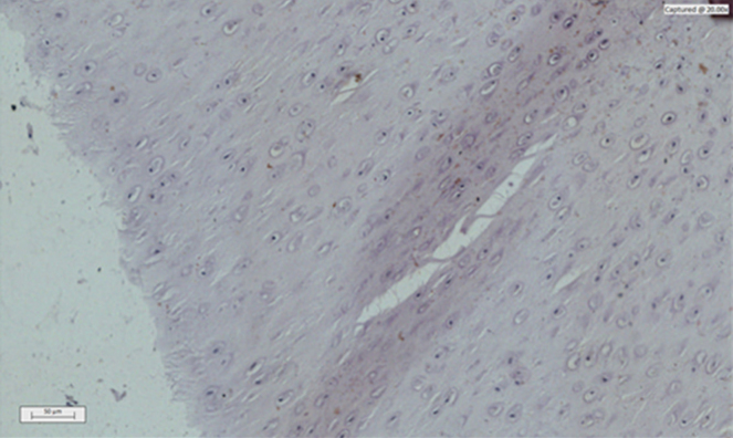
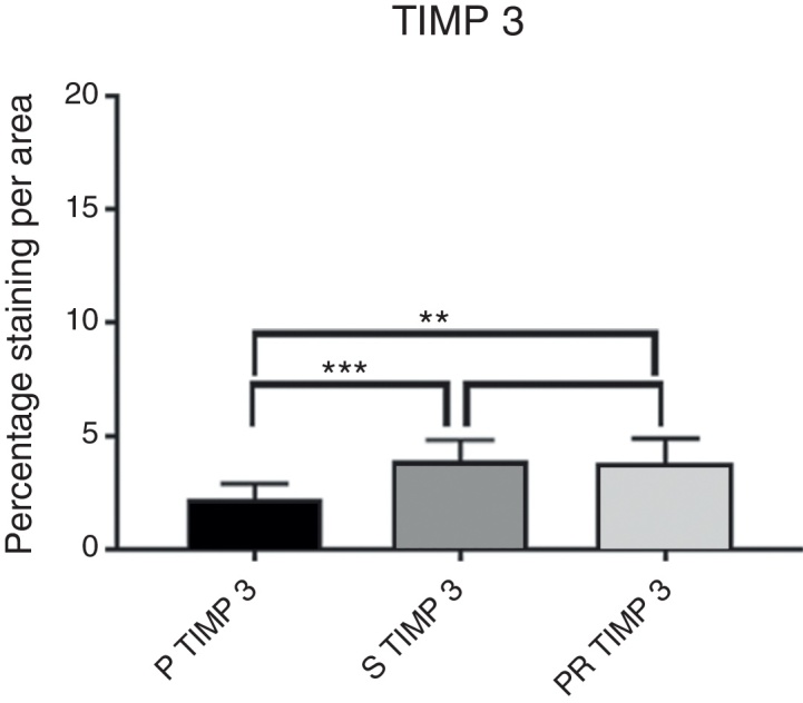
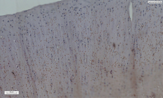
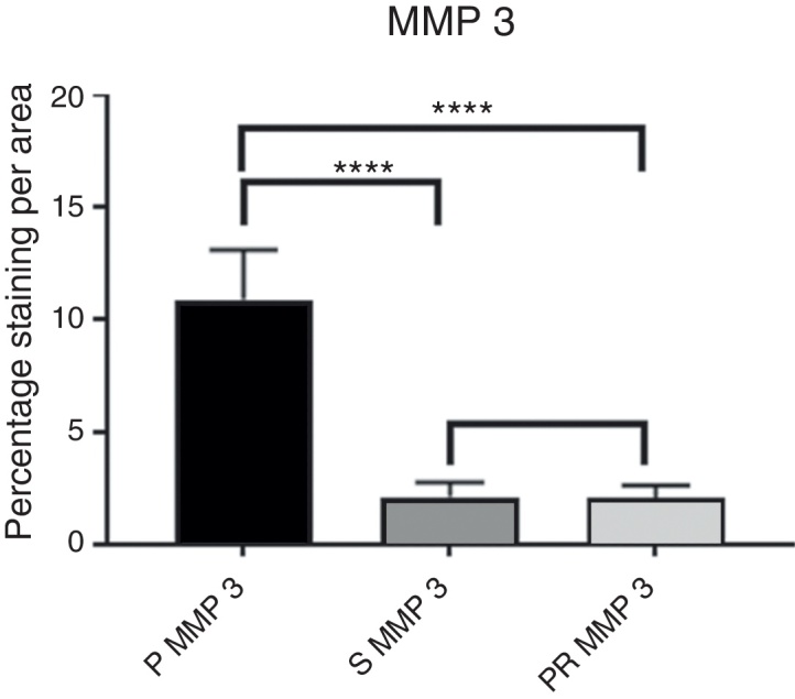
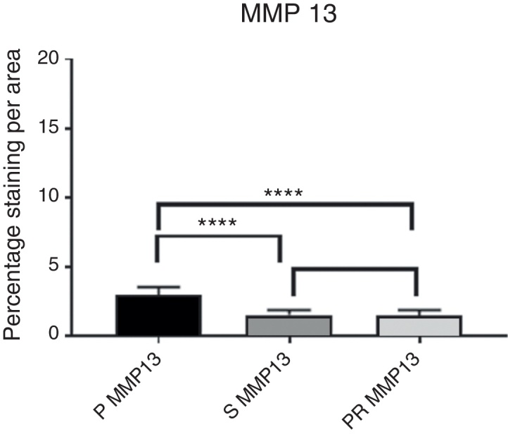
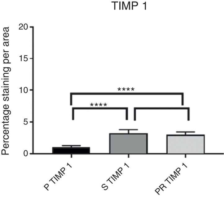
Forty-four male California rabbits were randomly assigned to three different groups (PR, S, and P) and submitted to the resection of the anterior cruciate ligament of the right knee. Three weeks after the surgical procedure, three intra-articular weekly injections were carried out with low-molecular-weight native hyaluronic acid (Hyalgan) to PR group, high molecular weight branched chain hyaluronic acid (Synvisc) to group S, and saline solution 0.9% to group P. All animals were sacrificed 12 weeks after the surgical procedure, and the tibial plateaus of the infiltrated knees were then dissected. Histological sections of cartilage from the tibial plateau support areas were stained with immunohistochemical markers in order to investigate the amount of metalloproteases (MMPs 3 and 13) and their inhibitors (TIMPs 1 and 3). The staining intensity was quantified on a Zeiss Imager.Z2 Metasystems microscope and analyzed by Metafer4 Msearch software.
The chondroprotective effect of the hyaluronic acids used in the study was demonstrated when compared to the control group. However, the comparison between them presented no significant statistical difference regarding chondroprotection.
The injection of saline solution demonstrated signs of OA development, while adding native hyaluronic acid of low molecular weight (Hyalgan) and hyaluronic acid of high molecular weight (Synvisc) protected the articular cartilage in this model of OA.
从免疫组织化学角度分析不同分子量透明质酸在兔骨关节炎实验模型中的作用。
44只雄性加利福尼亚兔随机分为三组(PR组、S组和P组),并对其右膝前交叉韧带进行切除。手术三周后,PR组每周进行三次关节内注射低分子量天然透明质酸(海乐妙),S组注射高分子量支链透明质酸(施沛特),P组注射0.9%生理盐水。所有动物在手术后12周处死,然后解剖注射侧膝关节的胫骨平台。对胫骨平台支撑区域软骨的组织学切片用免疫组织化学标记物染色,以研究金属蛋白酶(MMP-3和MMP-13)及其抑制剂(TIMP-1和TIMP-3)的含量。在蔡司Imager.Z2 Metasystems显微镜上对染色强度进行定量,并通过Metafer4 Msearch软件进行分析。
与对照组相比,本研究中使用的透明质酸具有软骨保护作用。然而,它们之间在软骨保护方面没有显著的统计学差异。
注射生理盐水显示出骨关节炎发展的迹象,而添加低分子量天然透明质酸(海乐妙)和高分子量透明质酸(施沛特)可在该骨关节炎模型中保护关节软骨。