Department of Medical Physics, University of Wisconsin, Madison, Wisconsin.
Department of Radiology, University of Wisconsin, Madison, Wisconsin.
Magn Reson Med. 2018 Dec;80(6):2586-2597. doi: 10.1002/mrm.27349. Epub 2018 Jun 12.
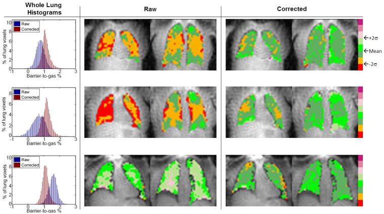
A novel technique is presented for retrospective estimation and removal of gas-phase hyperpolarized Xenon-129 (HP Xe) from images of HP Xe dissolved in the barrier (comprised of parenchymal lung tissue and blood plasma) and red blood cell (RBC) phases. The primary aim is mitigating RF pulse performance limitations on measures of gas exchange (e.g., barrier-gas and RBC-gas ratios). Correction for gas contamination would simplify technical dissemination of HP Xe applications across sites with varying hardware performance, scanner vendors, and models.
Digital lung phantom and human subject experiments (N = 8 healthy; N = 1 with idiopathic pulmonary fibrosis) were acquired with 3D radial trajectory and 1-point Dixon spectroscopic imaging to assess the correction method for mitigating barrier and RBC imaging artifacts. Dependence of performance on TE, image SNR, and gas contamination level were characterized. Inter- and intra-subject variation in the dissolved-phase ratios were quantified and compared to human subject experiments before and after correction.
Gas contamination resulted in image artifacts similar to those in disease that were mitigated after correction in both simulated and human subject data; for simulation experiments performance varied with TE, but was independent of image SNR and the amount of gas contamination. Artifacts and variation of barrier and RBC components were reduced after correction in both simulation and healthy human lungs (barrier, P = 0.01; RBC, P = 0.045).
The proposed technique significantly reduced regional variations in barrier and RBC ratios, separated using a 1-point Dixon approach, with improved accuracy of dissolved-phase HP Xe images confirmed in simulation experiments.
提出了一种从屏障相(由实质肺组织和血浆组成)和红细胞相(RBC)中溶解的超极化氙-129(HP Xe)的图像中回顾性估计和去除气相 HP Xe 的新技术。主要目的是减轻 RF 脉冲性能对气体交换测量(例如,屏障-气体和 RBC-气体比)的限制。气体污染的校正将简化 HP Xe 应用在具有不同硬件性能、扫描仪供应商和型号的不同站点之间的技术传播。
使用 3D 径向轨迹和 1 点 Dixon 光谱成像采集数字肺体模和人体实验(健康者 8 例,特发性肺纤维化 1 例),以评估校正方法对减轻屏障和 RBC 成像伪影的效果。评估了性能对 TE、图像 SNR 和气体污染水平的依赖性。定量评估和比较了溶解相比率的个体间和个体内变化,以及校正前后的人体实验。
气体污染导致与疾病相似的图像伪影,校正后在模拟和人体数据中均得到缓解;对于模拟实验,性能随 TE 变化,但与图像 SNR 和气体污染量无关。校正后,模拟和健康人体肺中的屏障和 RBC 成分的伪影和变化均减少(屏障,P=0.01;RBC,P=0.045)。
所提出的技术显著降低了使用 1 点 Dixon 方法分离的屏障和 RBC 比率的区域变化,在模拟实验中证实了溶解相 HP Xe 图像的准确性得到了提高。