Physical Sciences Platform, Sunnybrook Research Institute, Toronto, Ontario, Canada.
Department of Medical Biophysics, University of Toronto, Toronto, Ontario, Canada.
Theranostics. 2018 Apr 16;8(11):2909-2926. doi: 10.7150/thno.24911. eCollection 2018.
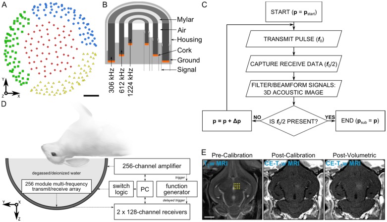
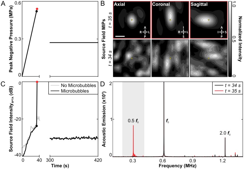
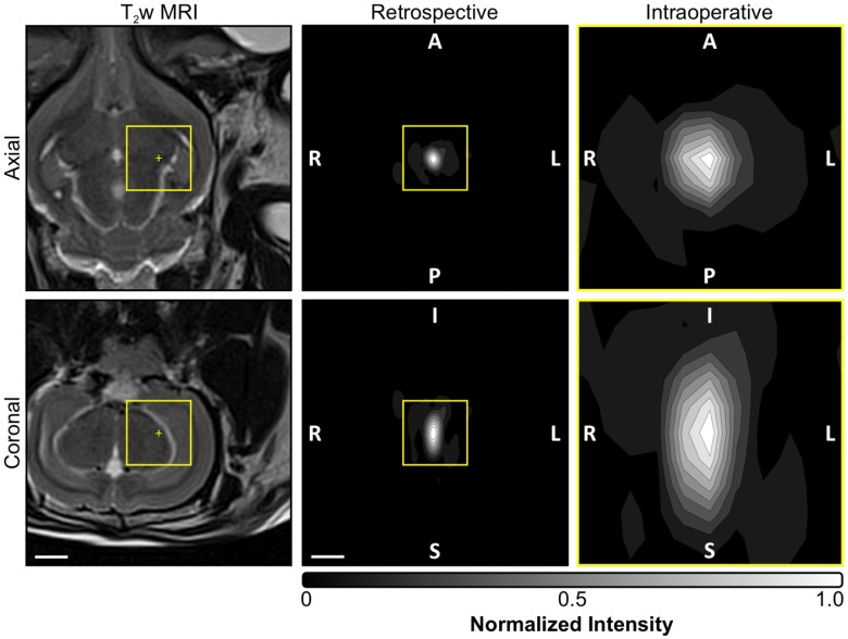
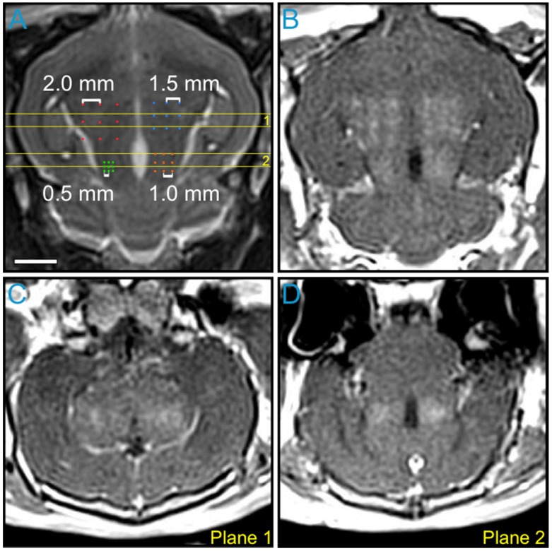
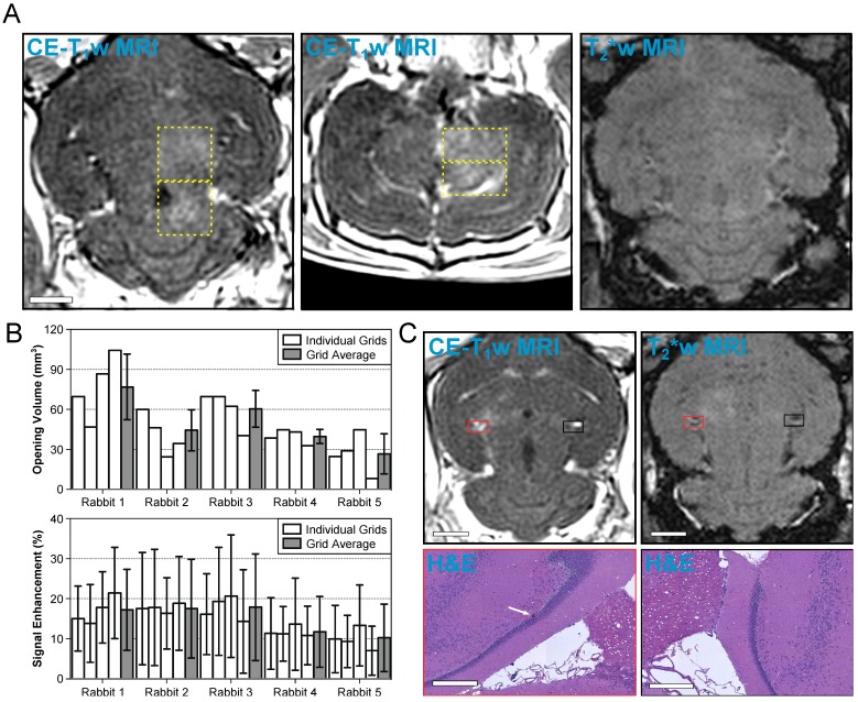
Focused ultrasound (FUS)-mediated blood-brain barrier (BBB) opening recently entered clinical testing for targeted drug delivery to the brain. Sources of variability exist in the current procedures, motivating the development of real-time monitoring and control techniques to improve treatment safety and efficacy. Here we used three-dimensional (3D) transcranial microbubble imaging to calibrate FUS exposure levels for volumetric BBB opening. Using a sparse hemispherical transmit/receive ultrasound phased array, pulsed ultrasound was focused transcranially into the thalamus of rabbits during microbubble infusion and multi-channel 3D beamforming was performed online with receiver signals captured at the subharmonic frequency. Pressures were increased pulse-by-pulse until subharmonic activity was detected on acoustic imaging (), and tissue volumes surrounding the calibration point were exposed at 50-100% via rapid electronic beam steering. Spatially-coherent subharmonic microbubble activity was successfully reconstructed transcranially during calibration sonications. Multi-point exposures induced volumetric regions of elevated BBB permeability assessed via contrast-enhanced magnetic resonance imaging (MRI). At exposure levels ≥75%, MRI and histological examination occasionally revealed tissue damage, whereas sonications at 50% were performed safely. Substantial intra-grid variability of FUS-induced bioeffects was observed via MRI, prompting future development of multi-point calibration schemes for improved treatment consistency. Receiver array sparsity and sensor configuration had substantial impacts on subharmonic detection sensitivity, and are factors that should be considered when designing next-generation clinical FUS brain therapy systems. Our findings suggest that 3D subharmonic imaging can be used to calibrate exposure levels for safe FUS-induced volumetric BBB opening, and should be explored further as a method for cavitation-mediated treatment guidance.
聚焦超声(FUS)介导的血脑屏障(BBB)开放最近进入了靶向递药到脑的临床测试。当前的操作程序中存在变异性来源,这促使开发实时监测和控制技术来提高治疗的安全性和有效性。在这里,我们使用三维(3D)经颅微泡成像来校准用于容积 BBB 开放的 FUS 暴露水平。使用稀疏的半球形发射/接收超声相控阵,在微泡输注期间将脉冲超声经颅聚焦到兔丘脑,并在线进行多通道 3D 波束形成,使用在次谐波频率下捕获的接收器信号进行。逐脉冲增加压力,直到在声学成像()上检测到次谐波活动,并且通过快速电子波束转向将校准点周围的组织体积暴露在 50-100%。在校准声处理期间成功地经颅重建了空间相干的次谐波微泡活动。多点暴露引起了通过对比增强磁共振成像(MRI)评估的 BBB 通透性升高的容积区域。在暴露水平≥75%时,MRI 和组织学检查偶尔会显示出组织损伤,而 50%的声处理则安全进行。通过 MRI 观察到 FUS 诱导的生物效应的网格内变异性很大,这促使为提高治疗一致性而开发多点校准方案。FUS 诱导的生物效应的接收阵列稀疏性和传感器配置对次谐波检测灵敏度有很大影响,在设计下一代临床 FUS 脑治疗系统时应考虑这些因素。我们的研究结果表明,3D 次谐波成像可用于校准安全的 FUS 诱导容积 BBB 开放的暴露水平,并且应该进一步探索作为空化介导的治疗指导方法。