Zhang Lin-Yuan, Lin Pan, Pan Jiaji, Ma Yuanyuan, Wei Zhenyu, Jiang Lu, Wang Liping, Song Yaying, Wang Yongting, Zhang Zhijun, Jin Kunlin, Wang Qian, Yang Guo-Yuan
1Department of Neurology, Ruijin Hospital, Shanghai Jiao Tong University School of Medicine, Shanghai, China.
2Medical Image Computing Lab and.
Aging Dis. 2018 Apr 1;9(2):262-272. doi: 10.14336/AD.2017.0613. eCollection 2018 Apr.
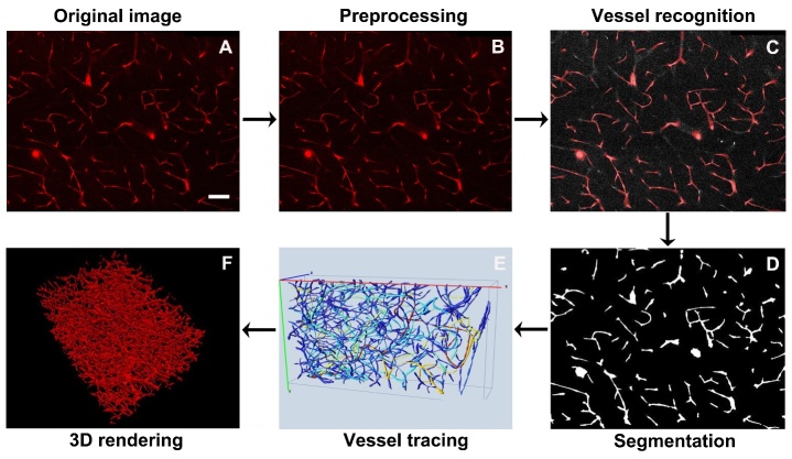
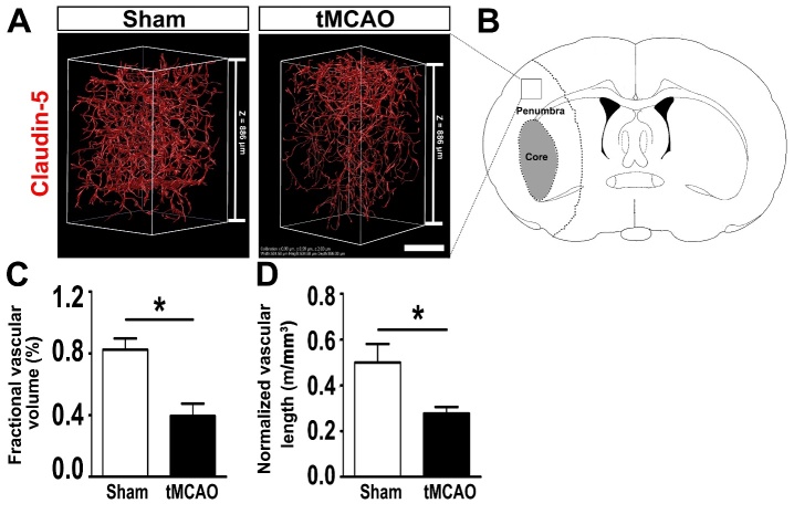
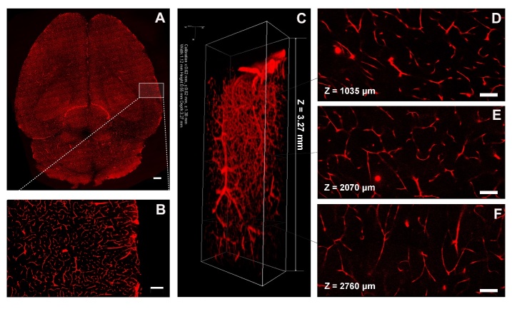
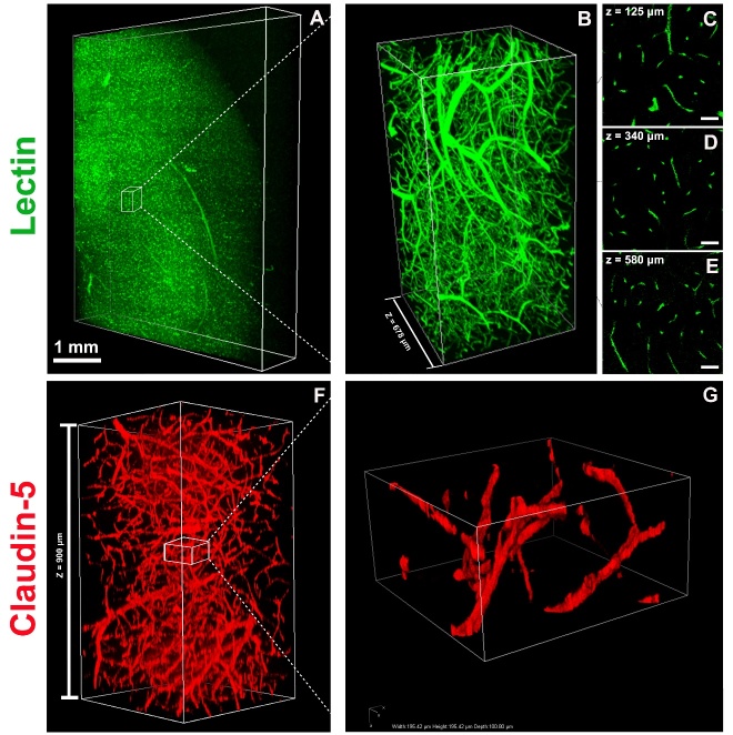
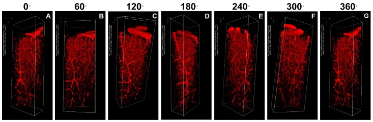
Elucidating the normal structure and distribution of cerebral vascular system is fundamental for understanding its function. However, studies on visualization and whole-brain quantification of vasculature with cellular resolution are limited. Here, we explored the structure of vasculature at the whole-brain level using the newly developed CLARITY technique. Adult male C57BL/6J mice undergoing transient middle cerebral artery occlusion and Tie2-RFP transgenic mice were used. Whole mouse brains were extracted for CLARITY processing. Immunostaining was performed to label vessels. Customized MATLAB code was used for image processing and quantification. Three-dimensional images were visualized using the Vaa3D software. Our results showed that whole mouse brain became transparent using the CLARITY method. Three-dimensional imaging and visualization of vasculature were achieved at the whole-brain level with a 1-μm voxel resolution. The quantitative results showed that the fractional vascular volume was 0.018 ± 0.004 mm per mm, the normalized vascular length was 0.44 ± 0.04 m per mm, and the mean diameter of the microvessels was 4.25 ± 0.08 μm. Furthermore, a decrease in the fractional vascular volume and a decrease in the normalized vascular length were found in the penumbra of ischemic mice compared to controls ( < 0.05). In conclusion, CLARITY provides a novel approach for mapping vasculature in the whole mouse brain at cellular resolution. CLARITY-optimized algorithms facilitate the assessment of structural change in vasculature after brain injury.
阐明脑血管系统的正常结构和分布是理解其功能的基础。然而,关于以细胞分辨率对脉管系统进行可视化和全脑定量分析的研究有限。在此,我们使用新开发的CLARITY技术探索了全脑水平的脉管系统结构。使用经历短暂大脑中动脉闭塞的成年雄性C57BL/6J小鼠和Tie2-RFP转基因小鼠。提取整个小鼠大脑进行CLARITY处理。进行免疫染色以标记血管。使用定制的MATLAB代码进行图像处理和定量分析。使用Vaa3D软件对三维图像进行可视化。我们的结果表明,使用CLARITY方法整个小鼠大脑变得透明。以1μm体素分辨率在全脑水平实现了脉管系统的三维成像和可视化。定量结果显示,血管分数体积为每毫米0.018±0.004立方毫米,归一化血管长度为每毫米0.44±0.04米,微血管平均直径为4.25±0.08μm。此外,与对照组相比,在缺血小鼠的半暗带中发现血管分数体积减少和归一化血管长度减少(<0.05)。总之,CLARITY提供了一种以细胞分辨率绘制整个小鼠大脑脉管系统的新方法。CLARITY优化算法有助于评估脑损伤后脉管系统的结构变化。