Division of Biomaterials and Biomechanics, Department of Restorative Dentistry, School of Dentistry, Oregon Health & Science University, Portland, OR, USA.
Biomedical Engineering Department, Knight Cancer Institute, OHSU Center for Spatial Systems Biomedicine, Oregon Health & Science University, Portland, OR, USA.
Sci Rep. 2019 Jul 26;9(1):10860. doi: 10.1038/s41598-019-47221-5.
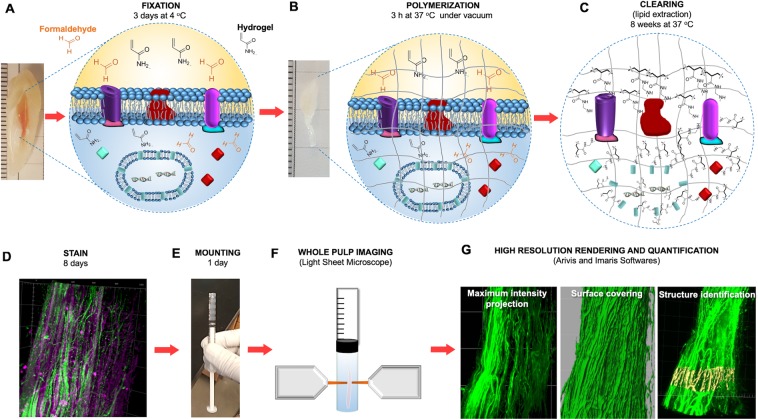
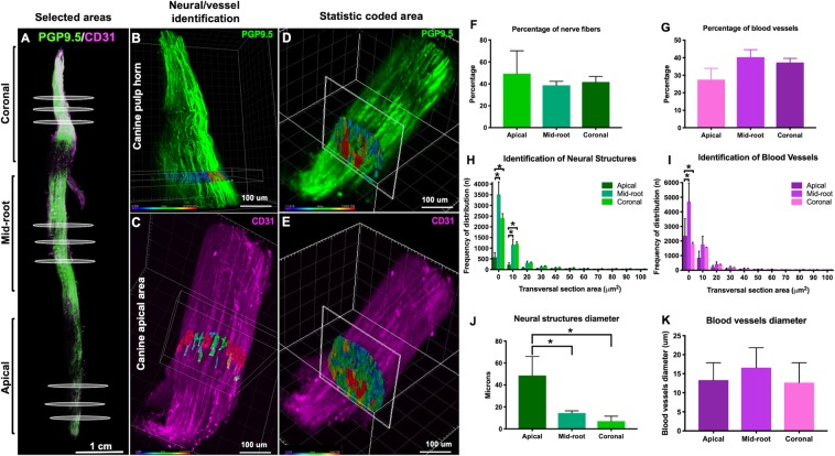
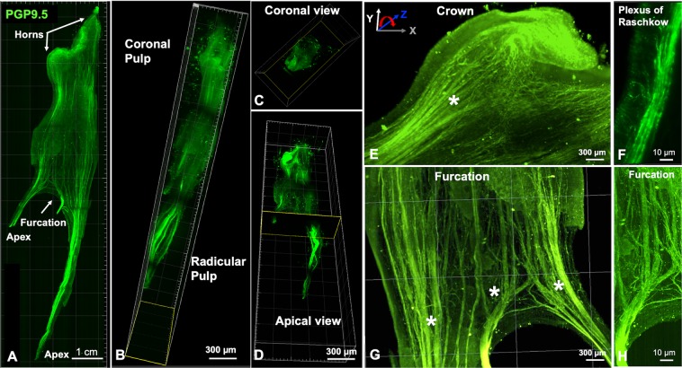
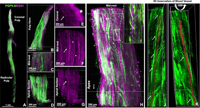
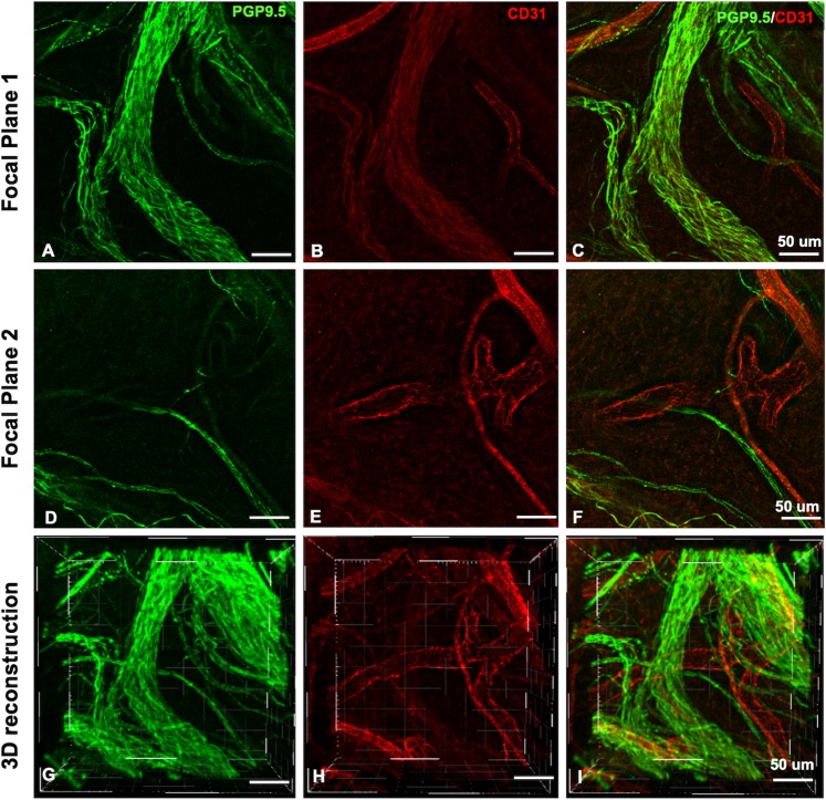
Direct visualization of the spatial relationships of the dental pulp tissue at the whole-organ has remained challenging. CLARITY (Clear Lipid-exchanged Acrylamide Tissue hYdrogel) is a tissue clearing method that has enabled successful 3-dimensional (3D) imaging of intact tissues with high-resolution and preserved anatomic structures. We used CLARITY to study the whole human dental pulp with emphasis on the neurovascular components. Dental pulps from sound teeth were CLARITY-cleared, immunostained for PGP9.5 and CD31, as markers for peripheral neurons and blood vessels, respectively, and imaged with light sheet microscopy. Visualization of the whole dental pulp innervation and vasculature was achieved. Innervation comprised 40% of the dental pulp volume and the vasculature another 40%. Marked innervation morphological differences between uni- and multiradicular teeth were found, also distinct neurovascular interplays. Quantification of the neural and vascular structures distribution, diameter and area showed that blood vessels in the capillary size range was twice as high as that of nerve fibers. In conclusion whole CLARITY-cleared dental pulp samples revealed 3D-morphological neurovascular interactions that could not be visualized with standard microscopy. This represents an outstanding tool to study the molecular and structural intricacies of whole dental tissues in the context of disease and treatment methods.
直接观察牙髓组织在整个器官中的空间关系一直具有挑战性。CLARITY(透明脂质交换丙烯酰胺组织水凝胶)是一种组织透明化方法,它能够成功地对完整组织进行高分辨率和保留解剖结构的三维(3D)成像。我们使用 CLARITY 研究了整个人类牙髓,重点研究了神经血管成分。从健康牙齿中提取牙髓,使用 CLARITY 进行透明化处理,然后分别用 PGP9.5 和 CD31 免疫染色,作为外周神经元和血管的标志物,并用光片显微镜进行成像。实现了整个牙髓神经支配和脉管系统的可视化。神经支配占牙髓体积的 40%,血管系统占 40%。发现单根和多根牙齿之间的神经支配形态存在明显差异,也存在明显的神经血管相互作用。对神经和血管结构的分布、直径和面积进行定量分析表明,毛细血管大小范围内的血管是神经纤维的两倍。总之,整个 CLARITY 透明化牙髓样本揭示了三维形态的神经血管相互作用,这是标准显微镜无法观察到的。这是研究整个牙髓组织在疾病和治疗方法背景下的分子和结构复杂性的一个杰出工具。