• 文献检索
  • 文档翻译
  • 深度研究
  • 学术资讯
  • Suppr Zotero 插件Zotero 插件
  • 邀请有礼
  • 套餐&价格
  • 历史记录
应用&插件
Suppr Zotero 插件Zotero 插件浏览器插件Mac 客户端Windows 客户端微信小程序
定价
高级版会员购买积分包购买API积分包
服务
文献检索文档翻译深度研究API 文档MCP 服务
关于我们
关于 Suppr公司介绍联系我们用户协议隐私条款
关注我们

Suppr 超能文献

核心技术专利:CN118964589B侵权必究
粤ICP备2023148730 号-1Suppr @ 2026

文献检索

告别复杂PubMed语法,用中文像聊天一样搜索,搜遍4000万医学文献。AI智能推荐,让科研检索更轻松。

立即免费搜索

文件翻译

保留排版,准确专业,支持PDF/Word/PPT等文件格式,支持 12+语言互译。

免费翻译文档

深度研究

AI帮你快速写综述,25分钟生成高质量综述,智能提取关键信息,辅助科研写作。

立即免费体验

小关节肥大是一个误称:一项回顾性研究。

Facet joint hypertrophy is a misnomer: A retrospective study.

作者信息

An Sang Joon, Seo Mi Sook, Choi Soo Il, Lim Tae-Ha, Shin So Jin, Kang Keum Nae, Kim Young Uk

机构信息

Department of Neurology Department of Anesthesiology and Pain Medicine, Catholic Kwandong University of Korea College of Medicine, International ST. Mary's Hospital, Incheon Department of Anesthesiology and Pain Medicine, Eulji General Hospital, Eulji University College of Medicine Department of Anesthesiology and Pain Medicine, National Police Hospital, Seoul, Republic of Korea.

出版信息

Medicine (Baltimore). 2018 Jun;97(24):e11090. doi: 10.1097/MD.0000000000011090.

DOI:10.1097/MD.0000000000011090
PMID:29901623
原文链接:https://pmc.ncbi.nlm.nih.gov/articles/PMC6024170/
Abstract

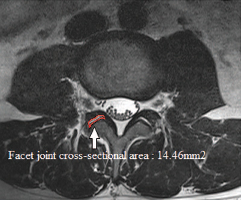
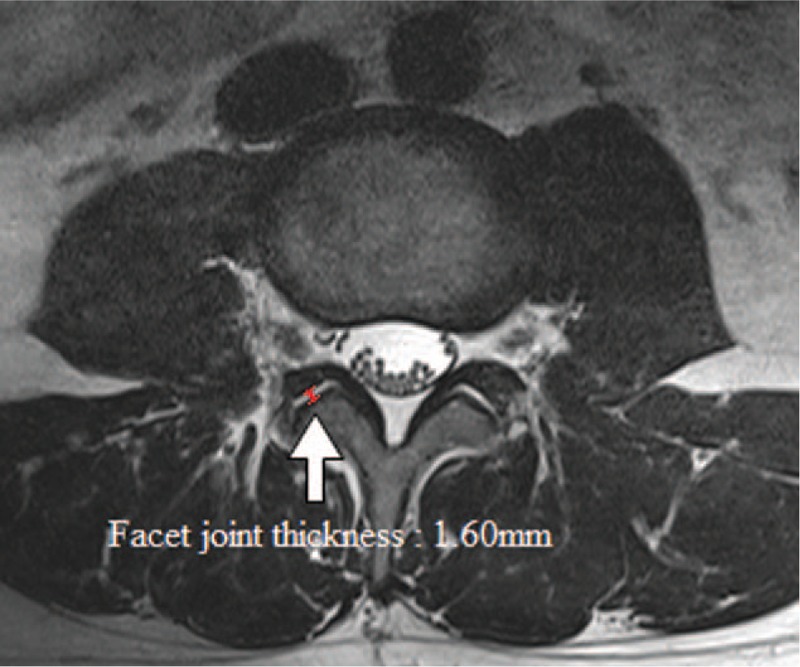
One of the major causes of lumbar spinal canal stenosis (LSCS) has been considered facet joint hypertrophy (FJH). However, a previous study asserted that "FJH" is a misnomer because common facet joints are no smaller than degenerative facet joints; however, this hypothesis has not been effectively demonstrated. Therefore, in order to verify that FJH is a misnomer in patients with LSCS, we devised new morphological parameters that we called facet joint thickness (FJT) and facet joint cross-sectional area (FJA).We collected FJT and FJA data from 114 patients with LSCS. A total of 86 control subjects underwent lumbar magnetic resonance imaging (MRI) as part of routine medical examinations, and axial T2-weighted MRI images were obtained from all participants. We measured FJT by drawing a line along the facet area and then measuring the narrowest point at L4-L5. We measured FJA as the whole cross-sectional area of the facet joint at the stenotic L4-L5 level.The average FJT was 1.60 ± 0.36 mm in the control group and 1.11 ± 0.32 mm in the LSCS group. The average FJA was 14.46 ± 5.17 mm in the control group and 9.31 ± 3.47 mm in the LSCS group. Patients with LSCS had significantly lower FJTs (P < .001) and FJAs (P < .001).FJH, a misnomer, should be renamed facet joint area narrowing. Using this terminology would eliminate confusion in descriptions of the facet joint.

摘要

腰椎管狭窄症(LSCS)的主要病因之一一直被认为是小关节肥大(FJH)。然而,先前的一项研究断言,“FJH”是一个误称,因为正常小关节并不比退变小关节小;然而,这一假设尚未得到有效证实。因此,为了验证FJH在LSCS患者中是一个误称,我们设计了新的形态学参数,即小关节厚度(FJT)和小关节截面积(FJA)。我们收集了114例LSCS患者的FJT和FJA数据。共有86名对照受试者作为常规体检的一部分接受了腰椎磁共振成像(MRI)检查,并从所有参与者中获取了轴向T2加权MRI图像。我们通过沿着小关节区域画一条线,然后测量L4-L5处最窄点来测量FJT。我们将FJA测量为狭窄的L4-L5水平处小关节的整个截面积。对照组的平均FJT为1.60±0.36mm,LSCS组为1.11±0.32mm。对照组的平均FJA为14.46±5.17mm,LSCS组为9.31±3.47mm。LSCS患者的FJT(P<0.001)和FJA(P<0.001)显著更低。FJH这个误称应重新命名为小关节面积缩小。使用这个术语将消除小关节描述中的混淆。

https://cdn.ncbi.nlm.nih.gov/pmc/blobs/776e/6024170/9aa579cfa38e/medi-97-e11090-g003.jpg
https://cdn.ncbi.nlm.nih.gov/pmc/blobs/776e/6024170/a74428bbaa0a/medi-97-e11090-g002.jpg
https://cdn.ncbi.nlm.nih.gov/pmc/blobs/776e/6024170/9aa579cfa38e/medi-97-e11090-g003.jpg
https://cdn.ncbi.nlm.nih.gov/pmc/blobs/776e/6024170/a74428bbaa0a/medi-97-e11090-g002.jpg
https://cdn.ncbi.nlm.nih.gov/pmc/blobs/776e/6024170/9aa579cfa38e/medi-97-e11090-g003.jpg

相似文献

1
Facet joint hypertrophy is a misnomer: A retrospective study.小关节肥大是一个误称:一项回顾性研究。
Medicine (Baltimore). 2018 Jun;97(24):e11090. doi: 10.1097/MD.0000000000011090.
2
Does lumbar facet fluid detected on magnetic resonance imaging correlate with radiographic instability in patients with degenerative lumbar disease?磁共振成像检测到的腰椎小关节积液与退行性腰椎疾病患者的影像学不稳定有关吗?
Spine (Phila Pa 1976). 2007 Jun 15;32(14):1555-60. doi: 10.1097/BRS.0b013e318067dc55.
3
Increased Facet Fluid Predicts Dynamic Changes in the Dural Sac Size on Axial-Loaded MRI in Patients with Lumbar Spinal Canal Stenosis.小关节液增多可预测腰椎管狭窄症患者轴向负荷MRI上硬脊膜囊大小的动态变化。
AJNR Am J Neuroradiol. 2016 Apr;37(4):730-5. doi: 10.3174/ajnr.A4582. Epub 2015 Nov 12.
4
Facet Sagittal Orientation: Possible Role in the Pathology of Degenerative Lumbar Spinal Stenosis.矢状面方位:在退行性腰椎管狭窄症病理学中的可能作用。
Spine (Phila Pa 1976). 2018 Jul 15;43(14):955-958. doi: 10.1097/BRS.0000000000002493.
5
Association between facet joint orientation/tropism and lumbar intervertebral disc degeneration.关节突关节方位/趋向与腰椎间盘退变的相关性。
Br J Neurosurg. 2024 Apr;38(2):293-300. doi: 10.1080/02688697.2020.1864289. Epub 2020 Dec 26.
6
Predictors of L4-L5 Degenerative Lumbar Spondylolisthesis: L4 Inclination Angle and Facet Joint Angle.L4-L5 退变性腰椎滑脱的预测因素:L4 倾斜角和关节突关节角。
World Neurosurg. 2019 Oct;130:e680-e686. doi: 10.1016/j.wneu.2019.06.188. Epub 2019 Jul 4.
7
Investigation of the Effect of Facet Joint Angle Characteristics on the Development of Lumbar Spinal Stenosis in Patients with Lumbar Canal Stenosis.探讨关节突关节角度特征对腰椎管狭窄症患者腰椎椎管狭窄发展的影响。
Acta Chir Orthop Traumatol Cech. 2023;90(6):383-390.
8
Congenital lumbar spinal stenosis: a prospective, control-matched, cohort radiographic analysis.先天性腰椎管狭窄症:一项前瞻性、对照匹配队列影像学分析。
Spine J. 2005 Nov-Dec;5(6):615-22. doi: 10.1016/j.spinee.2005.05.385.
9
Predictability of the effects of facet joint infiltration in the degenerate lumbar spine when assessing MRI scans.在评估MRI扫描时,退变腰椎小关节突关节浸润效果的可预测性。
J Orthop Surg Res. 2017 Nov 21;12(1):180. doi: 10.1186/s13018-017-0685-x.
10
The Role of the Ligamentum Flavum Area as a Morphological Parameter of Lumbar Central Spinal Stenosis.黄韧带区域作为腰椎中央椎管狭窄症形态学参数的作用
Pain Physician. 2017 Mar;20(3):E419-E424.

引用本文的文献

1
The Relationship between Neural Foraminal Stenosis and Imaging Features of Lumbar Spine MRI in Patients Older Than 60 Years with Lumbar Radiculopathy.60岁以上腰椎神经根病患者神经孔狭窄与腰椎MRI影像特征的关系
Taehan Yongsang Uihakhoe Chi. 2021 Jul;82(4):862-875. doi: 10.3348/jksr.2020.0095. Epub 2021 Apr 14.
2
Quantitative Radiological Characteristics of the Facet Joints in Patients with Lumbar Foraminal Stenosis.腰椎管狭窄症患者小关节的定量放射学特征
J Pain Res. 2022 Aug 17;15:2363-2371. doi: 10.2147/JPR.S374720. eCollection 2022.
3
In vivo relationships between lumbar facet joint and intervertebral disc composition and diurnal deformation.

本文引用的文献

1
Optimal Cut-Off Value of the Superior Articular Process Area as a Morphological Parameter to Predict Lumbar Foraminal Stenosis.作为预测腰椎椎间孔狭窄形态学参数的上关节突面积的最佳截断值
Pain Res Manag. 2017;2017:7914836. doi: 10.1155/2017/7914836. Epub 2017 Jan 9.
2
Biomechanical response of lumbar facet joints under follower preload: a finite element study.跟随者预载下腰椎小关节的生物力学响应:一项有限元研究。
BMC Musculoskelet Disord. 2016 Mar 15;17:126. doi: 10.1186/s12891-016-0980-4.
3
The nerve root sedimentation sign for differential diagnosis of lumbar spinal stenosis: a retrospective, consecutive cohort study.
腰椎小关节与椎间盘成分及昼夜变形的活体关系。
Clin Biomech (Bristol). 2021 Aug;88:105425. doi: 10.1016/j.clinbiomech.2021.105425. Epub 2021 Jul 14.
4
Percutaneous spinal endoscopy with unilateral interlaminar approach to perform bilateral decompression for central lumbar spinal stenosis: radiographic and clinical assessment.经皮脊柱内镜单侧椎板间入路双侧减压治疗中央型腰椎管狭窄症:影像学及临床评估
BMC Musculoskelet Disord. 2021 Mar 1;22(1):236. doi: 10.1186/s12891-021-04100-3.
神经根沉降征在腰椎管狭窄症鉴别诊断中的作用:一项回顾性、连续队列研究。
Eur Spine J. 2017 Oct;26(10):2512-2519. doi: 10.1007/s00586-016-4435-2. Epub 2016 Feb 13.
4
Epiduroscopic Removal of a Lumbar Facet Joint Cyst.经硬膜外腔镜下切除腰椎小关节囊肿。
Korean J Pain. 2015 Oct;28(4):275-9. doi: 10.3344/kjp.2015.28.4.275. Epub 2015 Oct 2.
5
Grading Osteoarthritic Changes of the Zygapophyseal Joints from Radiographs: A Reliability Study.通过X线片对关节突关节骨关节炎变化进行分级:一项可靠性研究。
J Manipulative Physiol Ther. 2015 Jun;38(5):344-51. doi: 10.1016/j.jmpt.2014.12.009. Epub 2015 Jun 26.
6
Measuring spinal canal size in lumbar spinal stenosis: description of method and preliminary results.测量腰椎管狭窄症中的椎管大小:方法描述及初步结果
Int J Spine Surg. 2015 Mar 24;9:3. doi: 10.14444/2008. eCollection 2015.
7
Characterization of symptomatic lumbar foraminal stenosis by conventional imaging.通过传统影像学对症状性腰椎管狭窄症的特征分析
Eur Spine J. 2015 Oct;24(10):2269-75. doi: 10.1007/s00586-015-3859-4. Epub 2015 Mar 15.
8
The anatomy and morphometry of cervical zygapophyseal joint meniscoids.颈椎关节突关节半月板的解剖学与形态测量学
Surg Radiol Anat. 2015 Sep;37(7):799-807. doi: 10.1007/s00276-014-1406-3. Epub 2014 Dec 20.
9
The prevalence of lumbar spine facet joint osteoarthritis and its association with low back pain in selected Korean populations.特定韩国人群中腰椎小关节骨关节炎的患病率及其与腰痛的关联。
Clin Orthop Surg. 2014 Dec;6(4):385-91. doi: 10.4055/cios.2014.6.4.385. Epub 2014 Nov 10.
10
Investigation of intervertebral disc and facet joint in lumbar spondylolisthesis using T2 mapping.利用T2映射技术对腰椎滑脱症中的椎间盘及小关节进行研究。
Magn Reson Med Sci. 2014;13(4):261-6. doi: 10.2463/mrms.2013-0099. Epub 2014 Oct 27.