Yoon Hye Eun, Kim Yaeni, Kim Sang Dong, Oh Jin Kyoung, Chung Yong-An, Shin Seok Joon, Yang Chul Woo, Seo Suk Min
Department of Internal Medicine, Incheon St. Mary's Hospital, Incheon, South Korea.
Division of Nephrology, Department of Internal Medicine, College of Medicine, The Catholic University of Korea, Seoul, South Korea.
Ann Transplant. 2018 Jun 15;23:412-421. doi: 10.12659/AOT.909212.
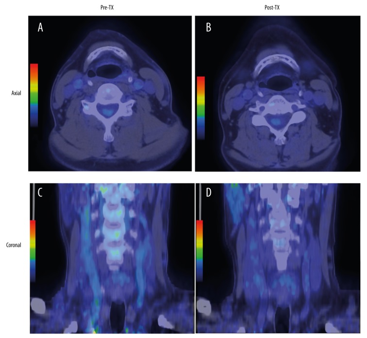
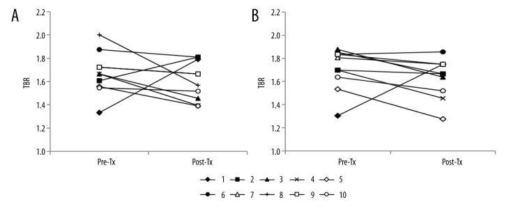
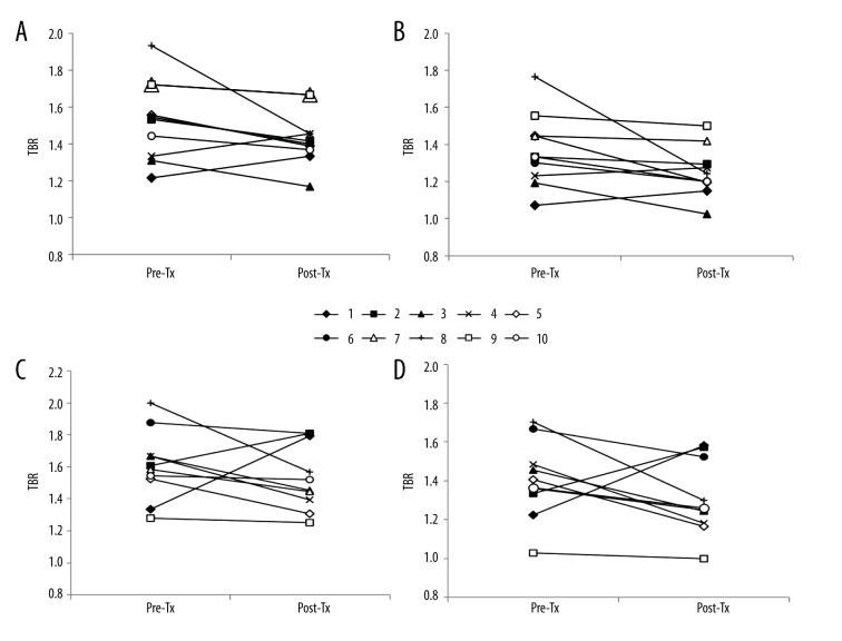
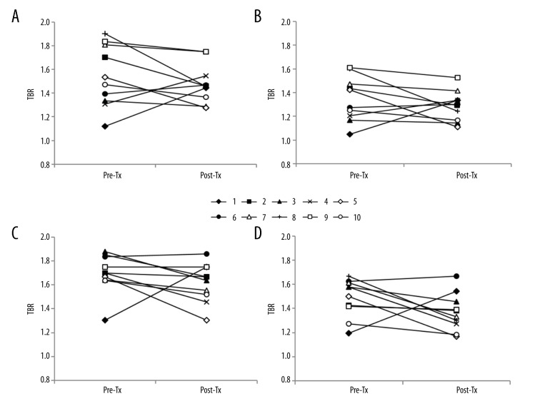
BACKGROUND Inflammatory activity of the artery can be assessed by measuring 18F-fluorodeoxyglucose (18F-FDG) uptake with positron emission tomography computed tomography (PET/CT). Improvement in vascular function after renal transplantation has been reported, but no studies have used 18F-FDG PET/CT to examine the changes in vascular inflammation. This study investigated the changes in the inflammatory activity in the carotid artery after renal transplantation in patients with chronic kidney disease (CKD). MATERIAL AND METHODS 18F-FDG PET/CT was performed before and at 4 months after transplantation. We quantified 18F-FDG uptake as the target-to-background ratio (TBR) in the carotid artery in 10 CKD patients. TBR was evaluated in the whole carotid artery (WH) and most-diseased segment (MDS), and the mean and maximum values were analyzed. The concentrations of inflammatory cytokines, including tumor necrosis factor-alpha, interleukin-6, plasminogen activator inhibitor-1, and endothelin-1, were measured. RESULTS Eight patients showed a decrease in mean or maximum TBR. The average mean or maximum TBRs in the WH and MDS of the right and left arteries were all reduced after transplantation. The average mean TBR for the right WH decreased significantly (% reduction [95% CI]) by -5.74% [-15.37, -0.02] (p=0.047). TBRs did not correlate significantly with cytokine concentrations. The changes in cytokine concentrations after transplantation varied. CONCLUSIONS 18F-FDG uptake by the WH and MDS tended to reduce after renal transplantation. Therefore, renal transplantation may confer an anti-inflammatory effect on carotid atherosclerosis in patients with CKD; however, this effect is not large enough to be demonstrated in this study with small sample size.
动脉的炎症活动可通过正电子发射断层扫描计算机断层扫描(PET/CT)测量18F-氟脱氧葡萄糖(18F-FDG)摄取来评估。已有报道称肾移植后血管功能有所改善,但尚无研究使用18F-FDG PET/CT来检查血管炎症的变化。本研究调查了慢性肾脏病(CKD)患者肾移植后颈动脉炎症活动的变化。材料与方法:在移植前及移植后4个月进行18F-FDG PET/CT检查。我们对10例CKD患者颈动脉中的18F-FDG摄取进行定量,以靶本比(TBR)表示。在整个颈动脉(WH)和病变最严重节段(MDS)评估TBR,并分析其平均值和最大值。测量包括肿瘤坏死因子-α、白细胞介素-6、纤溶酶原激活物抑制剂-1和内皮素-1在内的炎性细胞因子浓度。结果:8例患者的平均或最大TBR降低。移植后左右动脉WH和MDS的平均或最大TBR平均值均降低。右侧WH的平均TBR平均值显著降低(降低百分比[95%CI]),为-5.74%[-15.37,-0.02](p=0.047)。TBR与细胞因子浓度无显著相关性。移植后细胞因子浓度变化各异。结论:肾移植后WH和MDS的18F-FDG摄取趋于降低。因此,肾移植可能对CKD患者的颈动脉粥样硬化具有抗炎作用;然而,在本样本量较小的研究中,这种作用不够显著。