Health Research Institute, National Institute of Advanced Industrial Science and Technology (AIST), 2217-14, Hayashi-cho, Takamatsu, Kagawa, 761-0301, Japan.
Department of Biochemistry and Molecular Biology, Graduate School and Faculty of Medicine, The University of Tokyo, 7-3-1, Hongo, Bunkyo-ku, Tokyo, 113-0033, Japan.
Malar J. 2018 Jun 19;17(1):235. doi: 10.1186/s12936-018-2381-7.
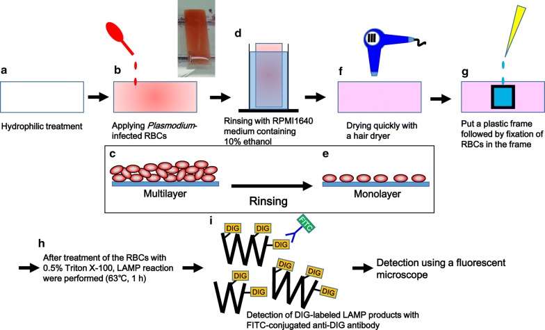
Five species of Plasmodium are known to infect humans. For proper treatment of malaria, accurate identification of the parasite species is crucial. The current gold standard for malaria diagnosis is microscopic examination of Giemsa-stained blood smears. Since the parasite species are identified by microscopists who manually search for the parasite-infected red blood cells (RBCs), misdiagnosis due to human error tends to occur in case of low parasitaemia or mixed infection. Then, molecular methods, such as polymerase chain reaction or loop-mediated isothermal amplification (LAMP), are required for conclusive identification of the parasite species. However, since molecular methods are highly sensitive, false-positive results tend to occur due to contamination (carry over) or the target gene products may be detected even after clearance of the parasites from the patient's blood. Therefore, accurate detection of parasites themselves by microscopic examination is essential for the definitive diagnosis. Thus, the method of in situ LAMP for the parasites was developed.
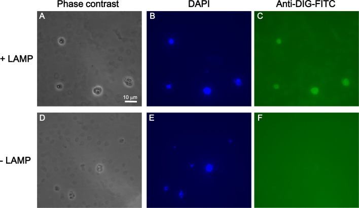
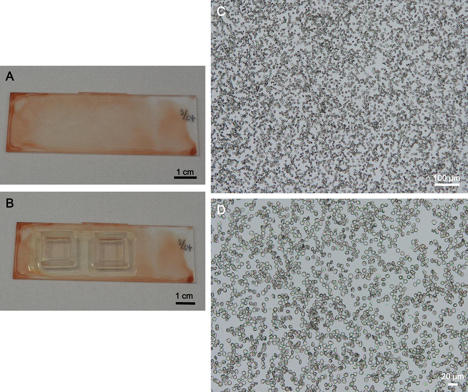
Red blood cell suspensions, including cultured Plasmodium falciparum, strain 3D7, infected-RBCs, were dispersed on cyclic olefin copolymer (COC) plate surfaces rendered hydrophilic by reactive ion-etching treatment using a SAMCO RIE system (hydrophilic-treated), followed by standing for 10 min to allow the RBCs to settle down on the plate surface. By rinsing the plate with RPMI 1640 medium, monolayers of RBCs formed on almost the entire plate surface. The plate was then dried with a hair drier. The RBCs were fixed with formalin, followed by permeabilization with Triton X-100. Then, amplification of the P. falciparum 18S rRNA gene by the LAMP reaction with digoxigenin (DIG)-labelled dUTP and a specific primer set was performed. Infected RBCs as fluorescence-positive cells with anti-DIG antibodies conjugated with fluorescein using fluorescent microscopy could be detected.
The present work shows that the potential of in situ LAMP for the identification of Plasmodium species at the single cell level on hydrophilic-treated COC palates, allowing highly sensitive and accurate malaria diagnosis. The findings will improve the efficacy of the gold standard method for malaria diagnosis.
已知有五种疟原虫可感染人类。为了正确治疗疟疾,准确识别寄生虫种类至关重要。目前,疟疾诊断的金标准是吉姆萨染色血涂片的显微镜检查。由于寄生虫种类是由显微镜检查者手动搜索寄生虫感染的红细胞(RBC)来识别的,因此在低寄生虫血症或混合感染的情况下,容易发生误诊。然后,需要聚合酶链反应或环介导等温扩增(LAMP)等分子方法来明确鉴定寄生虫种类。然而,由于分子方法高度敏感,由于污染(携带)或即使寄生虫从患者血液中清除后,也可能检测到靶基因产物,因此容易出现假阳性结果。因此,通过显微镜检查准确检测寄生虫本身对于明确诊断至关重要。因此,开发了寄生虫原位 LAMP 方法。
将包括培养的恶性疟原虫 3D7 株在内的红细胞悬浮液分散在经反应离子刻蚀处理的环烯烃共聚物(COC)板表面上,该处理使用 SAMCO RIE 系统进行(亲水处理),然后静置 10 分钟,使 RBC 在板表面沉降。通过用 RPMI 1640 培养基冲洗板,在几乎整个板表面形成 RBC 单层。然后用吹风机干燥该板。用福尔马林固定 RBC,然后用 Triton X-100 进行透化。然后,用 DIG 标记的 dUTP 和特定的引物组通过 LAMP 反应扩增恶性疟原虫 18S rRNA 基因。用抗 DIG 抗体与荧光素缀合的荧光显微镜检测感染 RBC 作为荧光阳性细胞。
本工作表明,在亲水处理的 COC 板上,原位 LAMP 具有在单细胞水平上识别疟原虫种类的潜力,从而实现高度敏感和准确的疟疾诊断。该发现将提高疟疾诊断金标准的效果。