Lee Shou-Wu, Lien Han-Chung, Lin Chi-Chen, Wen Mei-Chin, Chang Chi-Sen
Division of Gastroenterology, Department of Internal Medicine, Taichung Veterans General Hospital, Taichung, Taiwan.
Department of Internal Medicine, Chung Shan Medical University, Taichung, Taiwan.
Gastroenterology Res. 2018 Jun;11(3):189-194. doi: 10.14740/gr1009w. Epub 2018 May 31.
The aim of this study was to investigate the expression of transforming growth factor β (TGF-β) in the different stages of Barrett's esophagus (BE).
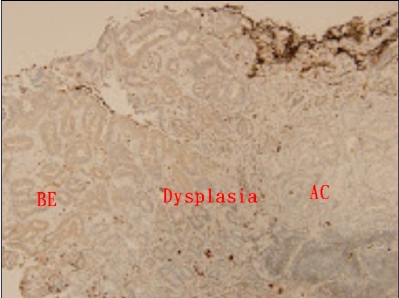
Paired endoscopic esophageal biopsy samples were obtained from patients with BE prospectively. Subjects were classified into three groups: BE, BE with dysplasia, and adenocarcinoma (AC) arising from BE. Biopsy specimens over normal esophageal epithelium and gastric cardiac epithelium of limited cases were done. Four cell lines, HETA1 (human esophageal epithelium), CA-A and CP-C (non-dysplastic metaplasia), and OE33 (AC) were analyzed for quantitative mRNA and Western blotting of TGF-β.
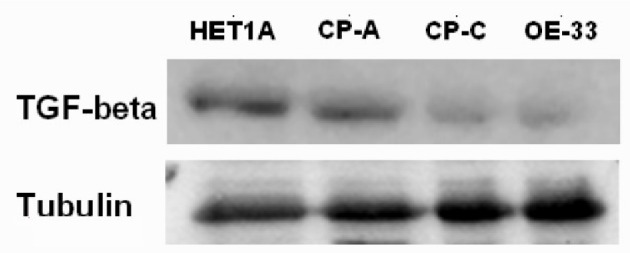
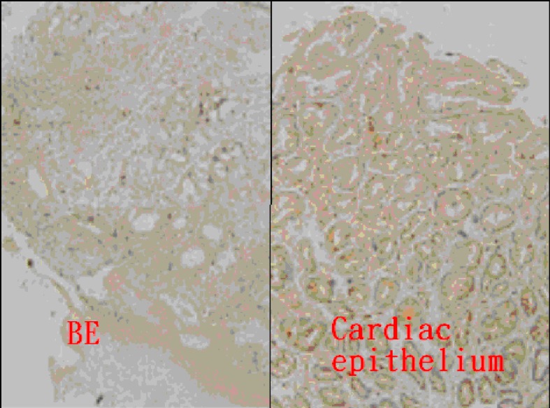
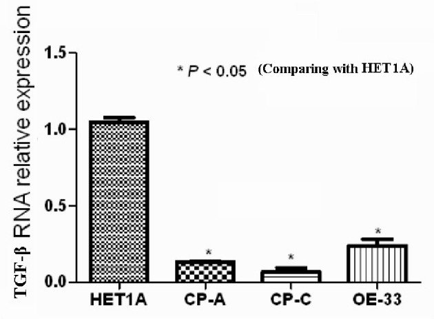
All 30 subjects with BE were enrolled. Expression of TGF-β mRNA in BE were significantly (P < 0.01) lower than that in the normal esophagus and cardiac epithelium. The BE tissue showed a lower positive ratio of TGF-β immunohistochemical (IHC) stain than the cardiac epithelium. The expression of TGF-β mRNA in the cell lines CA-A, CP-3, OE-33, was significantly (P < 0.05) lower than that in the cell line HETA-1. The Western blotting result showed lower TGF-β protein expression of the cell lines CA-A, CP-3, and OE-33.
The expression of TGF-β was lower in the tissue of BE.
本研究旨在调查转化生长因子β(TGF-β)在巴雷特食管(BE)不同阶段的表达情况。
前瞻性地从BE患者中获取配对的内镜食管活检样本。受试者分为三组:BE、伴有发育异常的BE和由BE引发的腺癌(AC)。对有限病例的正常食管上皮和胃贲门上皮进行活检标本检测。分析四种细胞系,即HETA1(人食管上皮)、CA-A和CP-C(非发育异常化生)以及OE33(AC)中TGF-β的定量mRNA和蛋白质印迹。
共纳入30例BE受试者。BE中TGF-β mRNA的表达显著低于正常食管和贲门上皮(P < 0.01)。BE组织中TGF-β免疫组化(IHC)染色的阳性率低于贲门上皮。细胞系CA-A、CP-3、OE-33中TGF-β mRNA的表达显著低于细胞系HETA-1(P < 0.05)。蛋白质印迹结果显示细胞系CA-A、CP-3和OE-33中TGF-β蛋白表达较低。
BE组织中TGF-β的表达较低。