College of Nursing, University of Iowa, Iowa City, Iowa.
School of Nursing, University of Wisconsin-Madison, Madison, Wisconsin.
Brain Behav. 2018 Jul;8(7):e01029. doi: 10.1002/brb3.1029. Epub 2018 Jun 19.
Sleep-disordered breathing is common in individuals with heart failure and may contribute to changes in the brain and decreased cognition. However, limited research has explored how the apnea-hypopnea index contributes to brain structure and cognition in this population. The aims of this study were to explore how the apnea-hypopnea index is associated with brain volume and cognition in heart failure patients.
Data of 28 heart failure patients (mean age = 67.93; SD = 5.78) were analyzed for this cross-sectional observational study. We evaluated the apnea-hypopnea index using a portable multichannel sleep-monitoring device. All participants were scanned using 3.0 Tesla magnetic resonance imaging and neuropsychological tests. Brain volume was evaluated using a voxel-based morphometry method with T1-weighted images. We used multiple regressions to analyze how the apnea-hypopnea index is associated with brain volume and cognition.
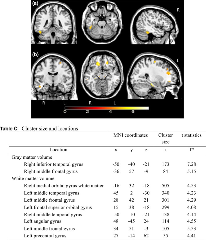
We found an inverse association between apnea-hypopnea index scores and white matter volume (β = -0.002, p = 0.026), but not in gray matter volume (β = -0.001, p = 0.237). Higher apnea-hypopnea index was associated with reduced regional gray and white matter volume (p < 0.001, uncorrected). Cognitive scores were not associated with the apnea-hypopnea index (p-values were >0.05).
Findings from this study provide exploratory evidence that higher apnea-hypopnea index may be associated with greater brain volume reduction in heart failure patients. Future studies are needed to establish the relationship between sleep-disordered breathing, brain volume, and cognition in heart failure samples.
睡眠呼吸障碍在心力衰竭患者中较为常见,可能导致大脑发生变化和认知能力下降。然而,关于睡眠呼吸暂停低通气指数(apnea-hypopnea index,AHI)在该人群中与大脑结构和认知功能的关系,目前研究较少。本研究旨在探讨心力衰竭患者的 AHI 与脑容量和认知功能的关系。
本横断面观察性研究分析了 28 例心力衰竭患者(平均年龄=67.93 岁,标准差=5.78 岁)的数据。我们使用便携式多导睡眠监测仪评估 AHI。所有参与者均接受 3.0 特斯拉磁共振成像和神经心理学测试。采用基于体素的形态测量学方法,使用 T1 加权图像评估脑容量。我们使用多元回归分析来探讨 AHI 与脑容量和认知功能的关系。
我们发现 AHI 评分与脑白质体积呈负相关(β=-0.002,p=0.026),但与脑灰质体积无相关性(β=-0.001,p=0.237)。较高的 AHI 与区域性灰质和白质体积减少相关(p<0.001,未校正)。认知评分与 AHI 无相关性(p 值均>0.05)。
本研究结果提供了探索性证据,表明较高的 AHI 可能与心力衰竭患者脑容量减少有关。需要进一步的研究来确定心力衰竭患者中睡眠呼吸障碍、脑容量和认知功能之间的关系。