• 文献检索
  • 文档翻译
  • 深度研究
  • 学术资讯
  • Suppr Zotero 插件Zotero 插件
  • 邀请有礼
  • 套餐&价格
  • 历史记录
应用&插件
Suppr Zotero 插件Zotero 插件浏览器插件Mac 客户端Windows 客户端微信小程序
定价
高级版会员购买积分包购买API积分包
服务
文献检索文档翻译深度研究API 文档MCP 服务
关于我们
关于 Suppr公司介绍联系我们用户协议隐私条款
关注我们

Suppr 超能文献

核心技术专利:CN118964589B侵权必究
粤ICP备2023148730 号-1Suppr @ 2026

文献检索

告别复杂PubMed语法,用中文像聊天一样搜索,搜遍4000万医学文献。AI智能推荐,让科研检索更轻松。

立即免费搜索

文件翻译

保留排版,准确专业,支持PDF/Word/PPT等文件格式,支持 12+语言互译。

免费翻译文档

深度研究

AI帮你快速写综述,25分钟生成高质量综述,智能提取关键信息,辅助科研写作。

立即免费体验

睾丸间质细胞瘤的治疗:一例报告。

Management of testicular Leydig cell tumor: A case report.

作者信息

Zhu Jianguo, Luan Yun, Li Haige

机构信息

Department of Radiology, the Second Affiliated Hospital of Nanjing Medical University Department of Ultrasound, Affiliated Hospital of Nanjing University of Traditional Chinese Medicine, Nanjing, China.

出版信息

Medicine (Baltimore). 2018 Jun;97(25):e11158. doi: 10.1097/MD.0000000000011158.

DOI:10.1097/MD.0000000000011158
PMID:29924022
原文链接:https://pmc.ncbi.nlm.nih.gov/articles/PMC6023788/
Abstract

RATIONALE

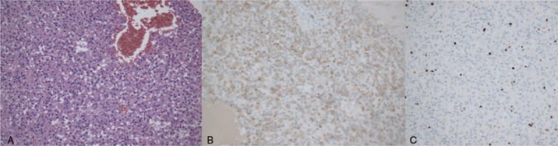
Testicular Leydig cell tumor (LCT) is a rare neoplasm. It commonly presents as a painless testicular mass with or without endocrine changes. Histological and immunohistochemical examination play important roles in differentiating LCT from testicular germ cell tumors.

PATIENT CONCERNS

We highlight the imaging phenotype, as well as the pathological findings of a case of LCT in a 62-year-old male.

DIAGNOSES

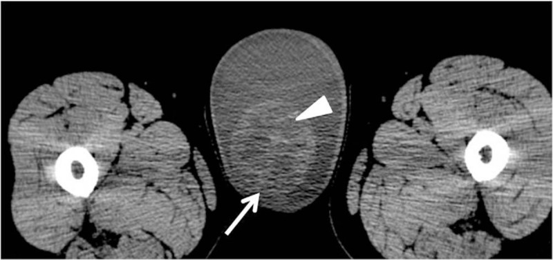
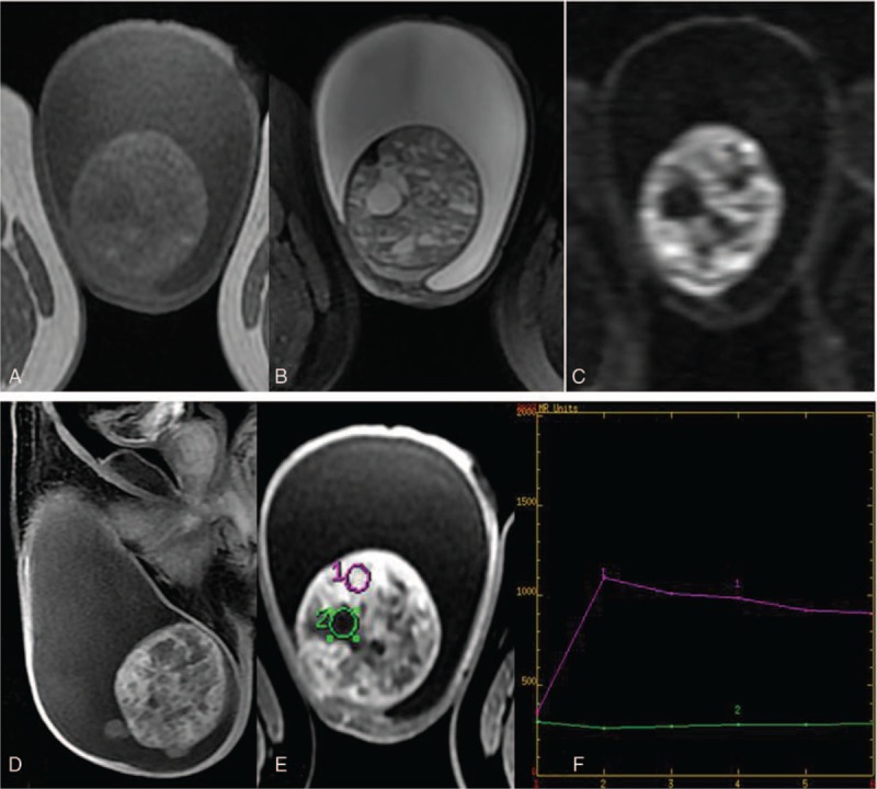
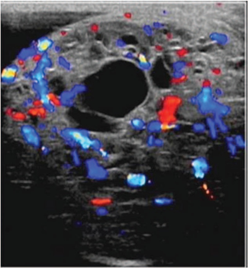
Preoperative noncontrast CT scan of the abdomen revealed a 7.0 × 6.4 × 5.3 cm oval mass with heterogeneous density, located in the right testis. Pelvic noncontrast MRI showed a heterogeneous mass on T1-weighted and T2-weighted images. The solid part of the tumor exhibited high signal on the diffusion-weighted imaging, and an obvious enhancement on the contrast-enhanced MR imaging. Ultrasonography examination demonstrated a large mixed echogenic space occupying lesion involving the whole right testis with multiple cystic areas and increased vascularity. This patient underwent radical orchiectomy. The pathologic diagnosis was LCT.

INTERVENTIONS

This patient underwent operative resection of the tumor. Due to the negative resection margins and absence of distant metastases, the patient did not receive additional radiotherapy or chemotherapy.

OUTCOMES

Four months after the surgery, the follow-up CT-scan did not reveal any local recurrence and distant metastases.

LESSONS

This case improves our ability to detect and diagnose LCT by summarizing its imaging characteristics as well as reviewing the literature. Additionally, we described the state-of-the-art management of the management of this rare tumor.

摘要

原理

睾丸间质细胞瘤(LCT)是一种罕见的肿瘤。它通常表现为无痛性睾丸肿块,伴有或不伴有内分泌改变。组织学和免疫组织化学检查在鉴别LCT与睾丸生殖细胞肿瘤方面发挥着重要作用。

患者情况

我们重点介绍了一名62岁男性LCT病例的影像学表现及病理结果。

诊断

术前腹部非增强CT扫描显示右侧睾丸有一个7.0×6.4×5.3 cm的椭圆形肿块,密度不均匀。盆腔非增强MRI在T1加权和T2加权图像上显示为不均匀肿块。肿瘤实性部分在扩散加权成像上呈高信号,在对比增强MRI上有明显强化。超声检查显示一个巨大的混合回声占位性病变累及整个右侧睾丸,有多个囊性区域且血管增多。该患者接受了根治性睾丸切除术。病理诊断为LCT。

干预措施

该患者接受了肿瘤手术切除。由于切缘阴性且无远处转移,患者未接受额外的放疗或化疗。

结果

术后4个月,随访CT扫描未发现任何局部复发和远处转移。

经验教训

通过总结其影像学特征并回顾文献,该病例提高了我们检测和诊断LCT的能力。此外,我们描述了这种罕见肿瘤的最新治疗方法。

https://cdn.ncbi.nlm.nih.gov/pmc/blobs/1a9f/6023788/3b793bb612b3/medi-97-e11158-g004.jpg
https://cdn.ncbi.nlm.nih.gov/pmc/blobs/1a9f/6023788/07ffa8f9b59c/medi-97-e11158-g001.jpg
https://cdn.ncbi.nlm.nih.gov/pmc/blobs/1a9f/6023788/5185fef3e224/medi-97-e11158-g002.jpg
https://cdn.ncbi.nlm.nih.gov/pmc/blobs/1a9f/6023788/fbd9ee5b9e45/medi-97-e11158-g003.jpg
https://cdn.ncbi.nlm.nih.gov/pmc/blobs/1a9f/6023788/3b793bb612b3/medi-97-e11158-g004.jpg

相似文献

1
Management of testicular Leydig cell tumor: A case report.睾丸间质细胞瘤的治疗:一例报告。
Medicine (Baltimore). 2018 Jun;97(25):e11158. doi: 10.1097/MD.0000000000011158.
2
Clinical presentation, management and follow-up of 83 patients with Leydig cell tumors of the testis: a prospective case-cohort study.睾丸间质细胞瘤 83 例患者的临床表现、治疗及随访:前瞻性病例队列研究。
Hum Reprod. 2019 Aug 1;34(8):1389-1403. doi: 10.1093/humrep/dez083.
3
Testicular tumors in children.儿童睾丸肿瘤
J Pediatr Surg. 2001 Dec;36(12):1796-801. doi: 10.1053/jpsu.2001.28841.
4
[The Leydig-cell testicular tumor. A case report with magnetic resonance images].[睾丸间质细胞瘤。一例伴有磁共振成像的病例报告]
Arch Esp Urol. 1998 Nov;51(9):928-31.
5
Ultrasound contrast-enhanced diagnosis of testicular Leydig cell tumor: A case report and literature review.超声造影增强诊断睾丸间质细胞瘤 1 例并文献复习
J Clin Ultrasound. 2024 Jun;52(5):643-648. doi: 10.1002/jcu.23665. Epub 2024 Mar 7.
6
Metastatic testicular germ cell tumor presenting with abdominal pain: CT and MRI findings.以腹痛为表现的转移性睾丸生殖细胞肿瘤:CT与MRI表现
JBR-BTR. 2009 Sep-Oct;92(5):256-8.
7
Surgery in infants and children with testicular and paratesticular tumours: a single centre experience over a 25-year-period.婴幼儿及儿童睾丸和睾丸旁肿瘤的手术治疗:25年单中心经验
Klin Padiatr. 2007 May-Jun;219(3):146-51. doi: 10.1055/s-2007-973847.
8
Leydig cell tumor of the testis with characteristic contrast patterns of tumor and non-tumorous testicular parenchyma on MRI: a case report.睾丸间质细胞瘤的 MRI 表现具有特征性的肿瘤和非肿瘤性睾丸实质对比模式:一例报告。
Abdom Radiol (NY). 2023 Aug;48(8):2477-2482. doi: 10.1007/s00261-023-03932-y. Epub 2023 May 10.
9
Leydig-cell tumour of the testis: retrospective analysis of clinical and therapeutic features in 204 cases.睾丸间质细胞瘤:204 例临床和治疗特征的回顾性分析。
World J Urol. 2020 Nov;38(11):2857-2862. doi: 10.1007/s00345-020-03079-1. Epub 2020 Jan 20.
10
Conservative surgical therapy for leydig cell tumor.睾丸间质细胞瘤的保守性手术治疗
J Urol. 2007 Aug;178(2):507-11; discussion 511. doi: 10.1016/j.juro.2007.03.108. Epub 2007 Jun 11.

引用本文的文献

1
Microdissection testicular sperm extraction for men with nonobstructive azoospermia who have a testicular tumor in situ at the time of sperm retrieval.对于在取精时存在原位睾丸肿瘤的非梗阻性无精子症男性,采用显微切割睾丸取精术。
Asian J Androl. 2025 May 1;27(3):423-427. doi: 10.4103/aja2024109. Epub 2025 Feb 28.
2
Successful management of Leydig Cell Tumor in a 65-year-Old patient: A rare case report.65岁患者睾丸间质细胞瘤的成功治疗:一例罕见病例报告
Urol Case Rep. 2025 Jan 2;58:102927. doi: 10.1016/j.eucr.2024.102927. eCollection 2025 Jan.
3
Testis sparing surgery for Leydig cell tumor, surgical approach, and clinical outcome: A case report and review of literature.

本文引用的文献

1
Giant mixed Sertoli-Leydig-Granulosa sex cord tumor of the testis; clinical, histopathological, and radiological features: a case report.睾丸巨大混合型支持-间质-颗粒性索肿瘤;临床、组织病理学及放射学特征:病例报告
Pan Afr Med J. 2017 May 19;27:51. doi: 10.11604/pamj.2017.27.51.10571. eCollection 2017.
2
Sertoli-Leydig Cell Tumor with Concurrent Rhabdomyosarcoma: Three Case Reports and a Review of the Literature.伴有横纹肌肉瘤的支持-间质细胞瘤:三例报告并文献复习
Case Rep Med. 2017;2017:4587296. doi: 10.1155/2017/4587296. Epub 2017 Jul 2.
3
Leydig Cell Tumor of Testis in a Child: An Uncommon Presentation.
睾丸支持细胞肿瘤的保留睾丸手术、手术方法及临床结果:一例病例报告并文献复习
SAGE Open Med Case Rep. 2024 May 27;12:2050313X241258365. doi: 10.1177/2050313X241258365. eCollection 2024.
4
Testicular leydig cell tumor revealed by hydrocele.鞘膜积液发现睾丸间质细胞瘤。
Urol Case Rep. 2020 Dec 5;35:101520. doi: 10.1016/j.eucr.2020.101520. eCollection 2021 Mar.
5
Differentiation of testicular seminoma and nonseminomatous germ cell tumor on magnetic resonance imaging.磁共振成像对睾丸精原细胞瘤和非精原细胞瘤性生殖细胞肿瘤的鉴别诊断
Medicine (Baltimore). 2019 Nov;98(45):e17937. doi: 10.1097/MD.0000000000017937.
儿童睾丸间质细胞瘤:一种罕见的表现。
J Indian Assoc Pediatr Surg. 2017 Jul-Sep;22(3):181-183. doi: 10.4103/jiaps.JIAPS_4_17.
4
Leydig cell tumor in grey zone: A case report.灰色地带的睾丸间质细胞瘤:一例报告
Int J Surg Case Rep. 2017;35:12-16. doi: 10.1016/j.ijscr.2017.03.043. Epub 2017 Apr 4.
5
[Metachronous Bilateral Testicular Tumors with Frequently Recurrent Hydrocele : A Case Report].[异时性双侧睾丸肿瘤伴频繁复发性鞘膜积液:病例报告]
Hinyokika Kiyo. 2017 Mar;63(3):115-118. doi: 10.14989/ActaUrolJap_63_3_115.
6
Atypical Leydig Cell Tumor in Children: Report of 2 Cases.儿童非典型性睾丸间质细胞瘤 2 例报告
Pediatrics. 2016 Nov;138(5). doi: 10.1542/peds.2016-0151.
7
Leydig cell tumor of the testis with azoospermia and elevated delta4 androstenedione: case report.睾丸间质细胞瘤伴无精子症及δ4雄烯二酮升高:病例报告
Basic Clin Androl. 2016 Nov 8;26:14. doi: 10.1186/s12610-016-0041-8. eCollection 2016.
8
Value of dynamic, contrast-enhanced MRI & intraoperative ultrasound for management of a nonpalpable, incidental, testicular Leydig-cell tumor.动态对比增强磁共振成像及术中超声在不可触及的偶然性睾丸间质细胞瘤管理中的价值
Radiol Case Rep. 2015 Nov 6;5(3):432. doi: 10.2484/rcr.v5i3.432. eCollection 2010.
9
Leydig cell tumor of testis with indeterminate features.具有不确定特征的睾丸间质细胞瘤。
Indian J Cancer. 2015 Oct-Dec;52(4):529-30. doi: 10.4103/0019-509X.178400.
10
Metachronous Bilateral Testicular Leydig-Like Tumors Leading to the Diagnosis of Congenital Adrenal Hyperplasia (Adrenogenital Syndrome).异时性双侧睾丸类Leydig细胞瘤导致先天性肾上腺增生(肾上腺生殖器综合征)的诊断。
Case Rep Pathol. 2015;2015:459318. doi: 10.1155/2015/459318. Epub 2015 Aug 16.