Department of Medicine, National Jewish Health, 1400 Jackson Street, Room A639, Denver, CO, 80206, USA.
Department of Biomedical Research and Center for Genes, Environment, and Health, National Jewish Health, University of Colorado, 1400 Jackson Street, Denver, CO, 80206, USA.
Respir Res. 2018 Jun 25;19(1):126. doi: 10.1186/s12931-018-0825-9.
Airway epithelial cells and alveolar macrophages (AMs) are the first line of defense in the lung during infection. Toll-like receptor (TLR) agonists have been extensively used to define the regulation of inflammation in these cells. However, previous studies were performed in non-paired airway epithelial cells and AMs. The major goal of our study was to compare the pro- and anti-inflammatory responses of paired human primary airway epithelial cells and AMs to TLR3 and TLR4 agonists.
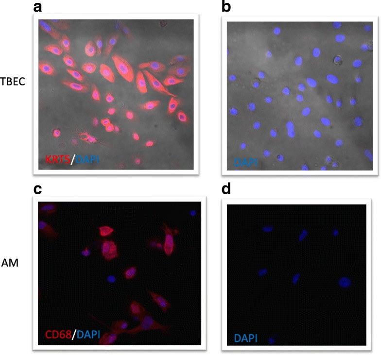
Tracheobronchial epithelial cells (TBEC) and AMs from four smokers and four non-smokers without lung disease were cultured with or without Poly(I:C) (PIC) (a TLR3 agonist) or LPS (a TLR4 agonist) for 4, 24 and 48 h. The immune responses of paired cells were compared.
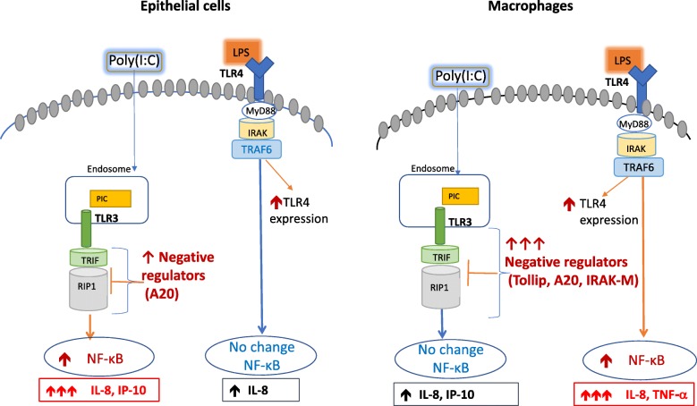
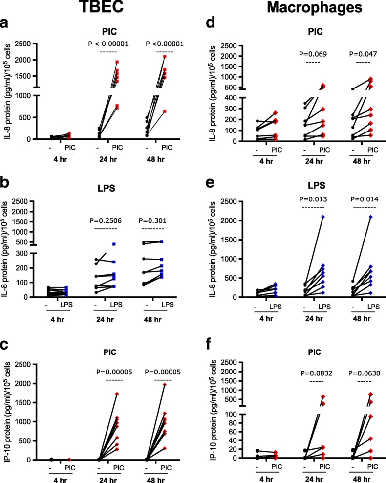
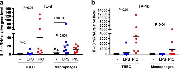
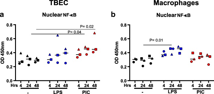
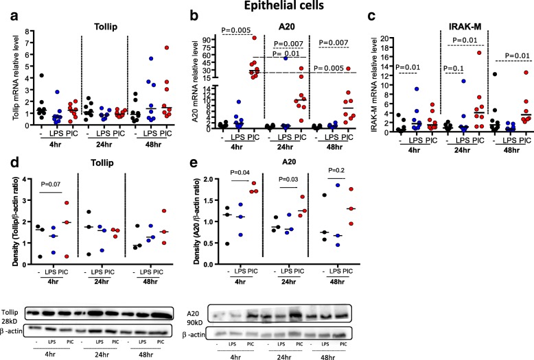
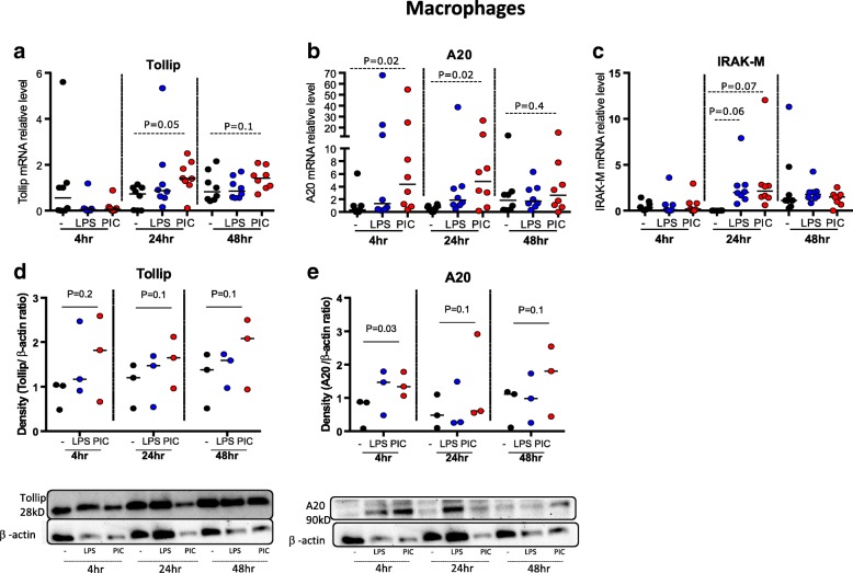
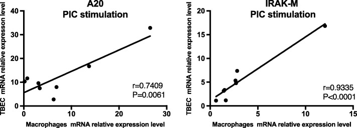
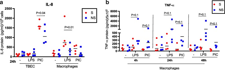
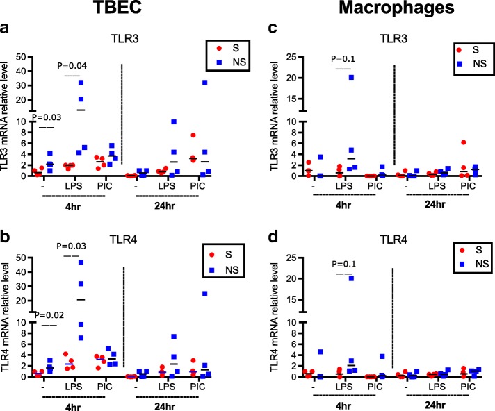
TBEC and AMs showed stronger pro-inflammatory cytokine (e.g., IL-8) responses to PIC and LPS, respectively. TLR3 and TLR4 mRNA levels were similar in non-stimulated TBEC and AMs. However, PIC stimulation in AMs led to sustained up-regulation of the immune negative regulators Tollip and A20, which may render AMs less sensitive to PIC stimulation than TBEC. Unlike AMs, TBEC did not increase NF-κB activation after LPS stimulation. Interestingly, smoking status was correlated with less TLR3 and IRAK-M expression in non-stimulated TBEC, but not in AMs. PIC-stimulated TBEC and LPS-stimulated AMs from smokers vs. non-smokers produced more IL-8. Finally, we show that expression of A20 and IRAK-M is strongly correlated in the two paired cell types.
By using paired airway epithelial cells and AMs, this study reveals how these two critical types of lung cells respond to viral and bacterial pathogen associated molecular patterns, and provides rationale for modulating immune negative regulators to prevent excessive lung inflammation during respiratory infection.
气道上皮细胞和肺泡巨噬细胞(AMs)是肺部感染时的第一道防线。Toll 样受体(TLR)激动剂已被广泛用于定义这些细胞中炎症的调节。然而,以前的研究是在非配对的气道上皮细胞和 AMs 中进行的。我们研究的主要目标是比较配对的人原代气道上皮细胞和 AMs 对 TLR3 和 TLR4 激动剂的促炎和抗炎反应。
从 4 名吸烟者和 4 名无肺部疾病的非吸烟者中培养气管支气管上皮细胞(TBEC)和 AMs,并用或不用 Poly(I:C)(TLR3 激动剂)或 LPS(TLR4 激动剂)培养 4、24 和 48 小时。比较配对细胞的免疫反应。
TBEC 和 AMs 对 PIC 和 LPS 的促炎细胞因子(如 IL-8)反应更强。未刺激的 TBEC 和 AMs 中的 TLR3 和 TLR4 mRNA 水平相似。然而,PIC 刺激 AMs 导致免疫负调节因子 Tollip 和 A20 的持续上调,这可能使 AMs 对 PIC 刺激的敏感性低于 TBEC。与 AMs 不同,TBEC 在用 LPS 刺激后不会增加 NF-κB 激活。有趣的是,吸烟状况与未刺激的 TBEC 中 TLR3 和 IRAK-M 的表达减少相关,但与 AMs 无关。与非吸烟者相比,吸烟者的 PIC 刺激的 TBEC 和 LPS 刺激的 AMs 产生更多的 IL-8。最后,我们表明 A20 和 IRAK-M 的表达在这两种配对细胞类型中强烈相关。
通过使用配对的气道上皮细胞和 AMs,本研究揭示了这两种关键类型的肺细胞如何对病毒和细菌病原体相关分子模式作出反应,并为调节免疫负调节因子以防止呼吸道感染期间过度的肺炎症提供了依据。