Turku PET Centre, University of Turku, Turku, Finland.
Turku PET Centre, Åbo Akademi University, Turku, Finland.
Sci Rep. 2018 Jun 26;8(1):9720. doi: 10.1038/s41598-018-27618-4.
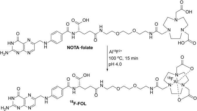
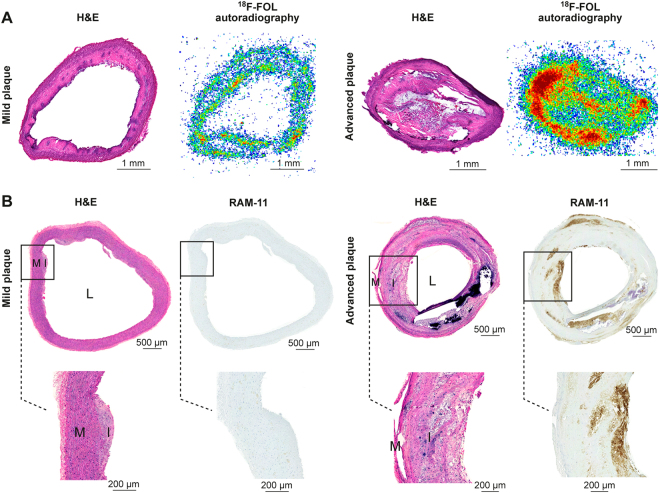
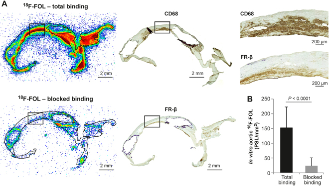
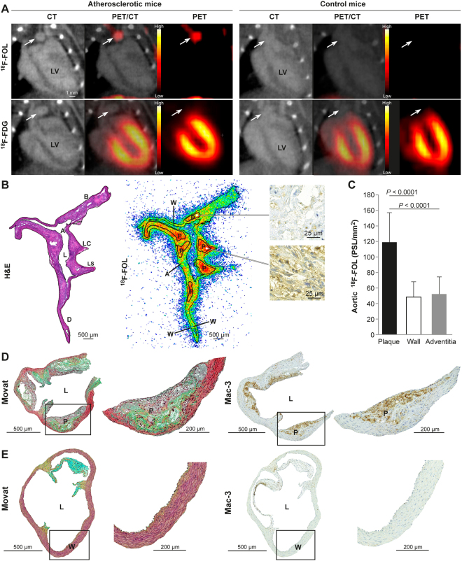
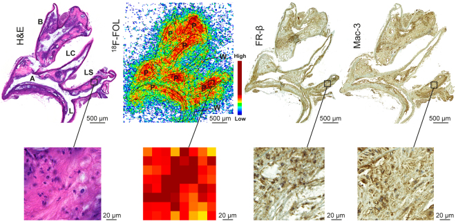
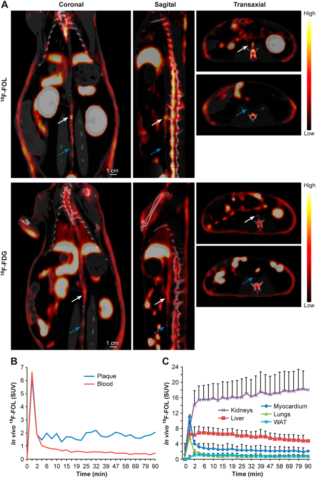
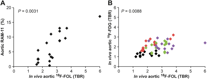
Inflammation plays an important role in the development of atherosclerosis and its complications. Because the folate receptor β (FR-β) is selectively expressed on macrophages, an FR targeted imaging agent could be useful for assessment of atherosclerotic inflammation. We investigated aluminum fluoride-18-labeled 1,4,7-triazacyclononane-1,4,7-triacetic acid conjugated folate (F-FOL) for the detection of atherosclerotic plaque inflammation. We studied atherosclerotic plaques in mice, rabbits, and human tissue samples using F-FOL positron emission tomography/computed tomography (PET/CT). Compound 2-deoxy-2-[F]fluoro-D-glucose (F-FDG) was used as a comparison. Firstly, we found that the in vitro binding of F-FOL co-localized with FR-β-positive macrophages in carotid endarterectomy samples from patients with recent ischemic symptoms. We then demonstrated specific accumulation of intravenously administered F-FOL in atherosclerotic plaques in mice and rabbits using PET/CT. We noticed that the F-FOL uptake correlated with the density of macrophages in plaques and provided a target-to-background ratio as high as F-FDG, but with considerably lower myocardial uptake. Thus, F-FOL PET/CT targeting of FR-β-positive macrophages presents a promising new tool for the in vivo imaging of atherosclerotic inflammation.
炎症在动脉粥样硬化及其并发症的发展中起着重要作用。由于叶酸受体 β(FR-β)选择性地在巨噬细胞上表达,因此 FR 靶向成像剂可用于评估动脉粥样硬化炎症。我们研究了用铝氟化物-18 标记的 1,4,7-三氮杂环壬烷-1,4,7-三乙酸偶联叶酸(F-FOL)用于检测动脉粥样硬化斑块炎症。我们使用 F-FOL 正电子发射断层扫描/计算机断层扫描(PET/CT)研究了小鼠、兔子和人组织样本中的动脉粥样硬化斑块。将 2-脱氧-2-[F]氟-D-葡萄糖(F-FDG)用作比较。首先,我们发现体外结合的 F-FOL 与来自近期有缺血症状的患者的颈动脉内膜切除术样本中的 FR-β阳性巨噬细胞共定位。然后,我们通过 PET/CT 证明了静脉内给予的 F-FOL 在小鼠和兔子的动脉粥样硬化斑块中的特异性积累。我们注意到,F-FOL 的摄取与斑块中巨噬细胞的密度相关,并提供了高达 F-FDG 的靶背比,但心肌摄取明显较低。因此,FR-β阳性巨噬细胞的 F-FOL PET/CT 靶向为动脉粥样硬化炎症的体内成像提供了一种很有前途的新工具。