CUO-Recherche, Centre de recherche du CHU de Québec and Département d'ophtalmologie, Faculté de médecine, Université Laval, Quebec, QC, Canada.
Department of Health Sciences and Technology, Brain Research Institute, University of Zurich, ETH Zurich, 8057, Zurich, Switzerland.
Cell Death Dis. 2018 Jun 27;9(7):727. doi: 10.1038/s41419-018-0780-x.
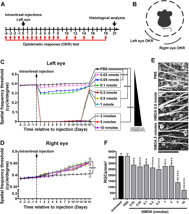
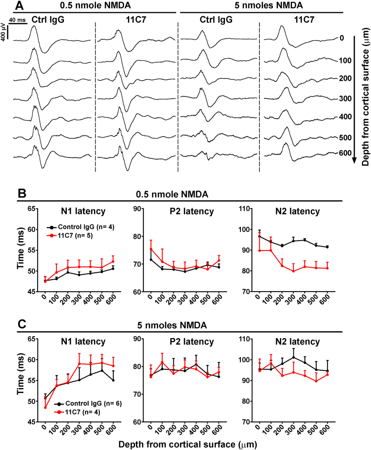
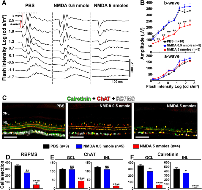
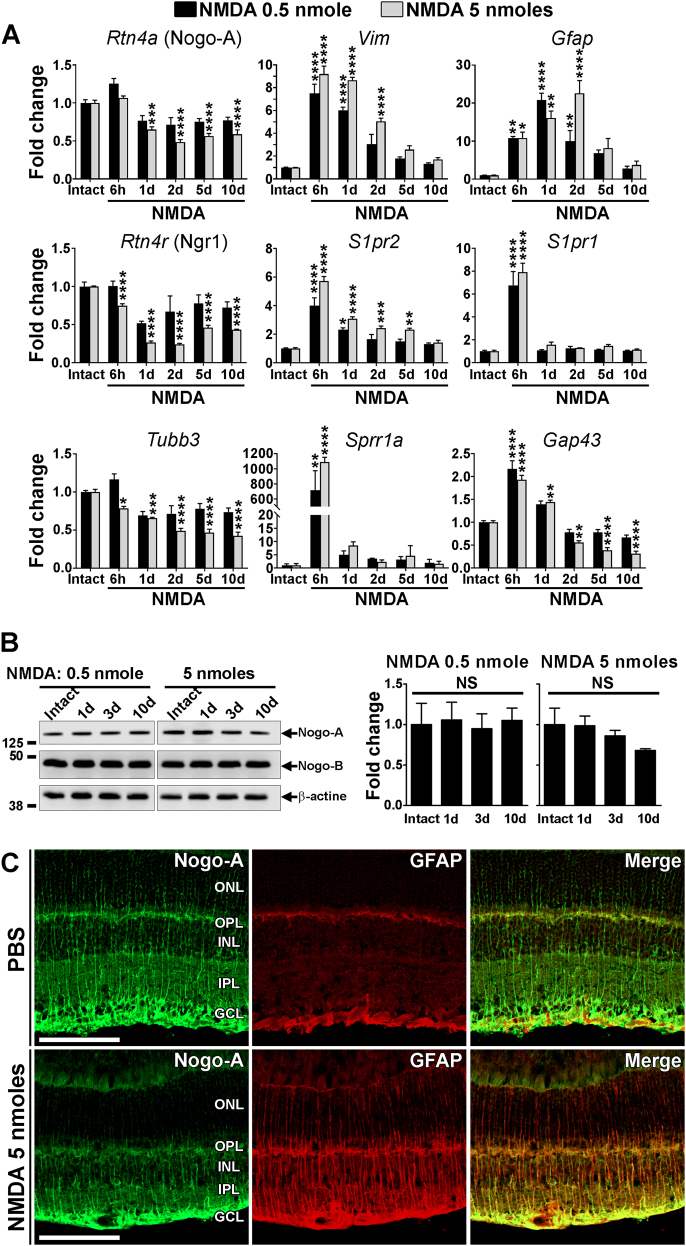
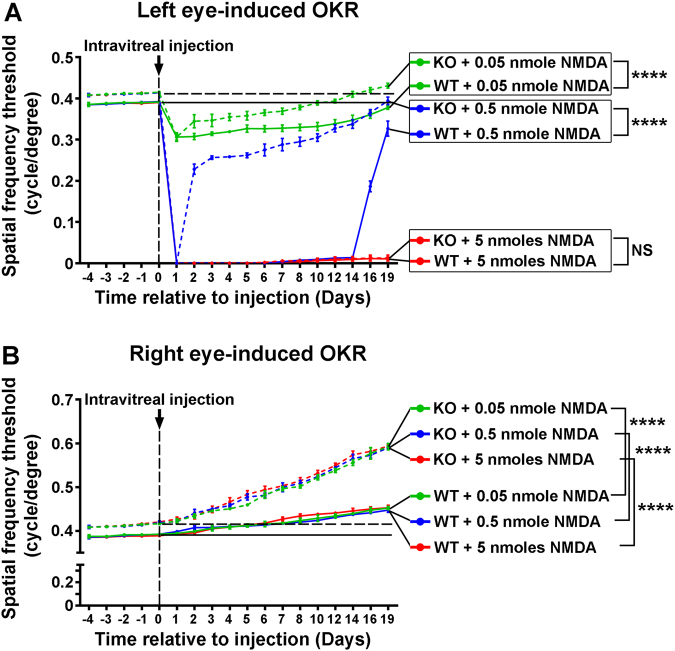
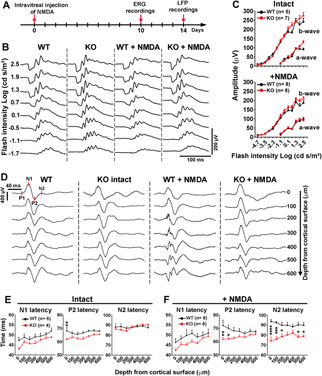
Myelin-associated proteins such as Nogo-A are major inhibitors of neuronal plasticity that contribute to permanent neurological impairments in the injured CNS. In the present study, we investigated the influence of Nogo-A on visual recovery after retinal injuries in mice. Different doses of N-methyl-D-aspartate (NMDA) were injected in the vitreous of the left eye to induce retinal neuron death. The visual function was monitored using the optokinetic response (OKR) as a behavior test, and electroretinogram (ERG) and local field potential (LFP) recordings allowed to assess changes in retinal and cortical neuron activity, respectively. Longitudinal OKR follow-ups revealed reversible visual deficits after injection of NMDA ≤ 1 nmole in the left eye and concomitant functional improvement in the contralateral visual pathway of the right eye that was let intact. Irreversible OKR loss observed with NMDA ≥ 2 nmol was correlated with massive retinal cell death and important ERG response decline. Strikingly, the OKR mediated by injured and intact eye stimulation was markedly improved in Nogo-A KO mice compared with WT animals, suggesting that the inactivation of Nogo-A promotes visual recovery and plasticity. Moreover, OKR improvement was associated with shorter latency of the N2 wave of Nogo-A KO LFPs relative to WT animals. Strikingly, intravitreal injection of anti-Nogo-A antibody (11C7) in the injured eye exerted positive effects on cortical LFPs. This study presents the intrinsic ability of the visual system to recover from NMDA-induced retinal injury and its limitations. Nogo-A neutralization may promote visual recovery in retinal diseases such as glaucoma.
髓鞘相关蛋白,如 Nogo-A,是神经元可塑性的主要抑制剂,导致中枢神经系统损伤后的永久性神经功能障碍。在本研究中,我们研究了 Nogo-A 对小鼠视网膜损伤后视觉恢复的影响。在左眼玻璃体内注射不同剂量的 N-甲基-D-天冬氨酸 (NMDA) 以诱导视网膜神经元死亡。使用视觉运动反应 (OKR) 作为行为测试监测视觉功能,视网膜电图 (ERG) 和局部场电位 (LFP) 记录分别评估视网膜和皮质神经元活动的变化。纵向 OKR 随访显示,在左眼注射 NMDA≤1nmole 后出现可逆性视觉缺陷,同时对侧未损伤的右眼视觉通路功能改善。用 NMDA≥2nmol 观察到的不可逆 OKR 丧失与大量视网膜细胞死亡和重要的 ERG 反应下降相关。引人注目的是,与 WT 动物相比,Nogo-A KO 小鼠受伤和未受伤眼刺激的 OKR 明显改善,表明 Nogo-A 的失活促进了视觉恢复和可塑性。此外,与 WT 动物相比,Nogo-A KO LFP 的 N2 波潜伏期更短。引人注目的是,损伤眼内注射抗 Nogo-A 抗体 (11C7) 对皮质 LFP 产生了积极影响。本研究揭示了视觉系统从 NMDA 诱导的视网膜损伤中恢复的内在能力及其局限性。Nogo-A 中和可能促进青光眼等视网膜疾病的视觉恢复。