Ocular Angiogenesis Group, Departments of Ophthalmology and Medical Biology, Amsterdam University Medical Centers, Academic Medical Center, Amsterdam, The Netherlands.
School of Pharmaceutical Sciences, University of Geneva, Geneva, Switzerland.
Angiogenesis. 2018 Nov;21(4):823-836. doi: 10.1007/s10456-018-9627-4. Epub 2018 Jun 27.
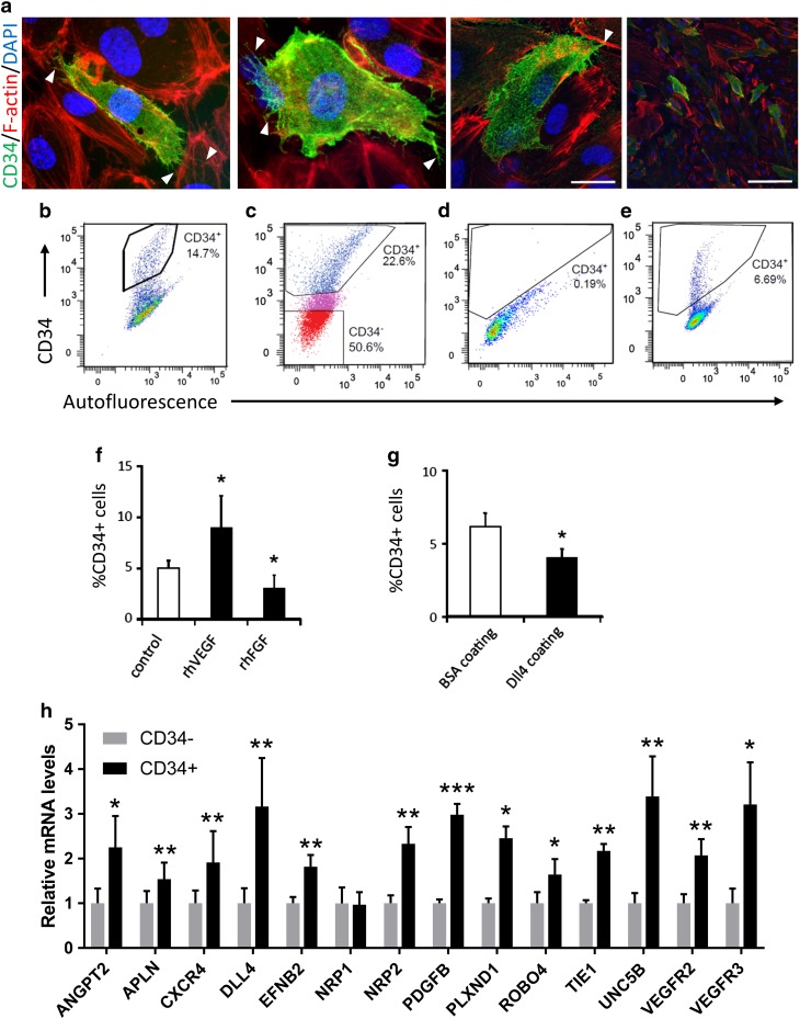
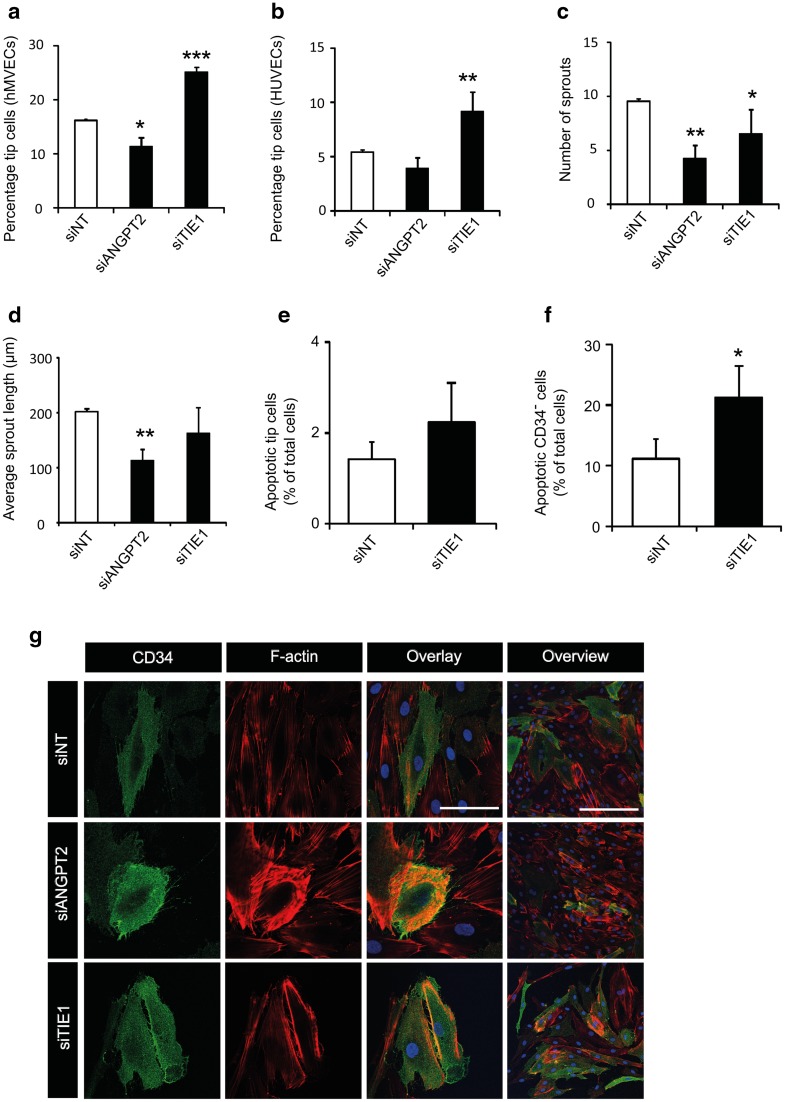
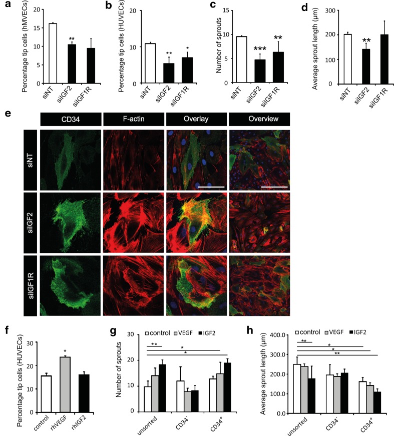
Tip cells, the leading cells of angiogenic sprouts, were identified in cultures of human umbilical vein endothelial cells (HUVECs) by using CD34 as a marker. Here, we show that tip cells are also present in primary human microvascular endothelial cells (hMVECs), a more relevant endothelial cell type for angiogenesis. By means of flow cytometry, immunocytochemistry, and qPCR, it is shown that endothelial cell cultures contain a dynamic population of CD34 cells with many hallmarks of tip cells, including filopodia-like extensions, elevated mRNA levels of known tip cell genes, and responsiveness to stimulation with VEGF and inhibition by DLL4. Furthermore, we demonstrate that our in vitro tip cell model can be exploited to investigate cellular and molecular mechanisms in tip cells and to discover novel targets for anti-angiogenesis therapy in patients. Small interfering RNA (siRNA) was used to knockdown gene expression of the known tip cell genes angiopoietin 2 (ANGPT2) and tyrosine kinase with immunoglobulin-like and EGF-like domains 1 (TIE1), which resulted in similar effects on tip cells and sprouting as compared to inhibition of tip cells in vivo. Finally, we identified two novel tip cell-specific genes in CD34 tip cells in vitro: insulin-like growth factor 2 (IGF2) and IGF-1-receptor (IGF1R). Knockdown of these genes resulted in a significant decrease in the fraction of tip cells and in the extent of sprouting in vitro and in vivo. In conclusion, this study shows that by using our in vitro tip cell model, two novel essential tip cells genes are identified.
通过使用 CD34 作为标志物,在人脐静脉内皮细胞(HUVEC)的培养物中鉴定出了血管生成芽的主导细胞——尖端细胞。在这里,我们表明尖端细胞也存在于原代人微血管内皮细胞(hMVEC)中,这是一种更相关的血管生成内皮细胞类型。通过流式细胞术、免疫细胞化学和 qPCR 表明,内皮细胞培养物中存在着一群具有许多尖端细胞特征的动态 CD34 细胞群体,包括类似纤毛的延伸、已知尖端细胞基因的 mRNA 水平升高,以及对 VEGF 的刺激和 DLL4 抑制的反应性。此外,我们证明我们的体外尖端细胞模型可用于研究尖端细胞中的细胞和分子机制,并发现针对患者抗血管生成治疗的新靶点。通过使用小干扰 RNA(siRNA)敲低已知的尖端细胞基因血管生成素 2(ANGPT2)和含免疫球蛋白样和表皮生长因子样结构域的酪氨酸激酶 1(TIE1)的基因表达,与体内抑制尖端细胞相比,这导致了尖端细胞和发芽的类似影响。最后,我们在体外的 CD34 尖端细胞中鉴定出两个新的尖端细胞特异性基因:胰岛素样生长因子 2(IGF2)和胰岛素样生长因子 1 受体(IGF1R)。这些基因的敲低导致体外和体内尖端细胞的比例和发芽程度显著降低。总之,这项研究表明,通过使用我们的体外尖端细胞模型,鉴定出了两个新的必需尖端细胞基因。