• 文献检索
  • 文档翻译
  • 深度研究
  • 学术资讯
  • Suppr Zotero 插件Zotero 插件
  • 邀请有礼
  • 套餐&价格
  • 历史记录
应用&插件
Suppr Zotero 插件Zotero 插件浏览器插件Mac 客户端Windows 客户端微信小程序
定价
高级版会员购买积分包购买API积分包
服务
文献检索文档翻译深度研究API 文档MCP 服务
关于我们
关于 Suppr公司介绍联系我们用户协议隐私条款
关注我们

Suppr 超能文献

核心技术专利:CN118964589B侵权必究
粤ICP备2023148730 号-1Suppr @ 2026

文献检索

告别复杂PubMed语法,用中文像聊天一样搜索,搜遍4000万医学文献。AI智能推荐,让科研检索更轻松。

立即免费搜索

文件翻译

保留排版,准确专业,支持PDF/Word/PPT等文件格式,支持 12+语言互译。

免费翻译文档

深度研究

AI帮你快速写综述,25分钟生成高质量综述,智能提取关键信息,辅助科研写作。

立即免费体验

流速、心率、血管几何形状和狭窄程度对冠状动脉血流动力学指标影响的数值分析。

Numerical analysis of the impact of flow rate, heart rate, vessel geometry, and degree of stenosis on coronary hemodynamic indices.

作者信息

Malota Zbigniew, Glowacki Jan, Sadowski Wojciech, Kostur Marcin

机构信息

Biocybernetics Laboratory, Prof. Z. Religa Foundation of Cardiac Surgery Development, Wolnosci st. 345a, 41-800, Zabrze, Poland.

Department of Radiology, Silesian Medical University, 3-go Maja st. 13/15, 41-800, Zabrze, Poland.

出版信息

BMC Cardiovasc Disord. 2018 Jun 28;18(1):132. doi: 10.1186/s12872-018-0865-6.

DOI:10.1186/s12872-018-0865-6
PMID:29954323
原文链接:https://pmc.ncbi.nlm.nih.gov/articles/PMC6025704/
Abstract

BACKGROUND

The stenosis of the coronary arteries is usually caused by atherosclerosis. Hemodynamic significance of patient-specific coronary stenoses and the risk of its progression may be assessed by comparing the hemodynamic effects induced by flow disorders. The present study shows how stenosis degree and variable flow conditions in coronary artery affect the oscillating shear index, residence time index, pressure drop coefficient and fractional flow reserve. We assume that changes in the hemodynamic indices in relation to variable flow conditions and geometries evaluated using the computational fluid dynamics may be an additional factor for a non-invasive assessment of the coronary stenosis detected on multi-slice computed tomography.

METHODS

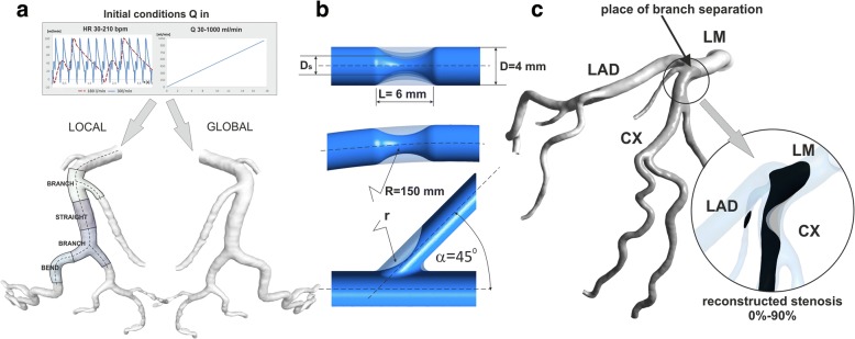
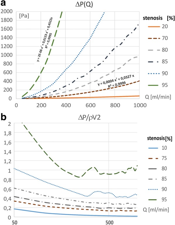
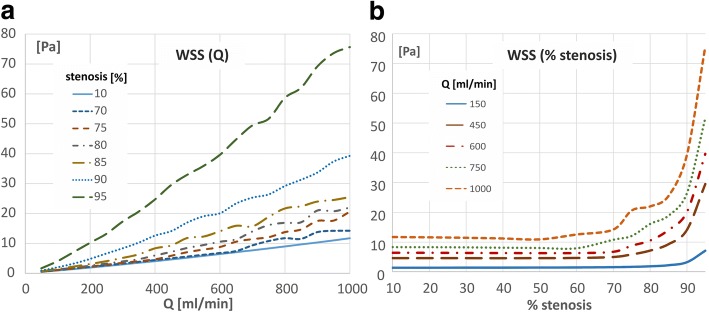
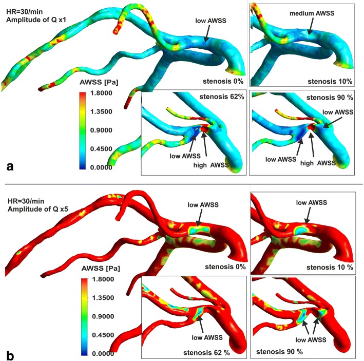
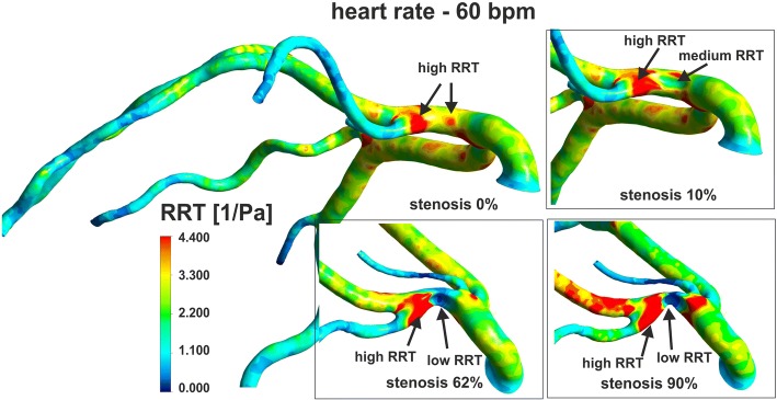
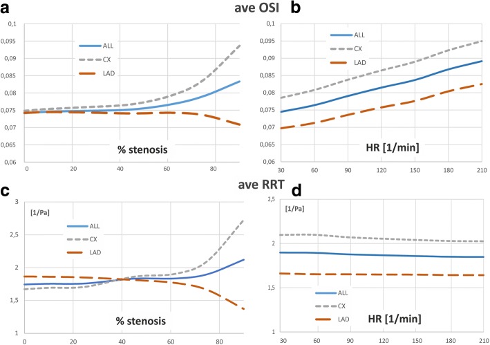
The local-parametrised models of basic shapes of the vessels, such as straight section, bend, and bifurcation as well as the global-patient-specific models of left coronary artery were used for numerical simulation of flow in virtually reconstructed stenotic vessels. Calculations were carried out for vessels both without stenosis, and vessels of 10 to 95% stenosis. The flow rate varied within the range of 20 to 1000 ml/min, and heart rate frequency within the range of 30 to 210 cycles/min. The computational fluid dynamics based on the finite elements method verified by the experimental measurements of the velocity profiles was used to analyse blood flow in the coronary arteries.

RESULTS

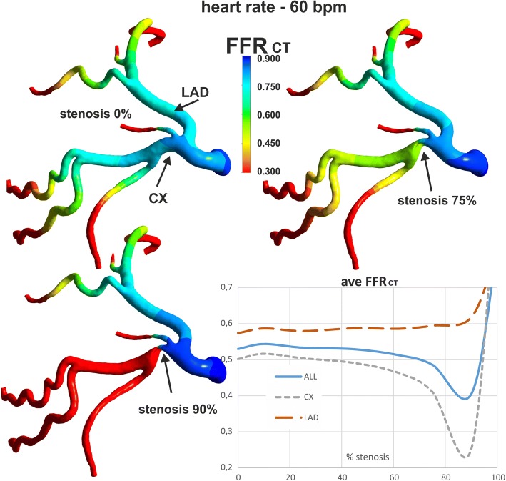
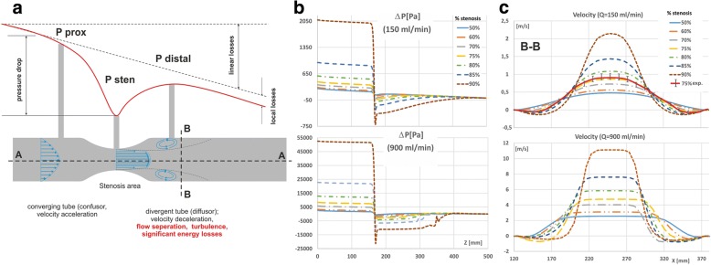
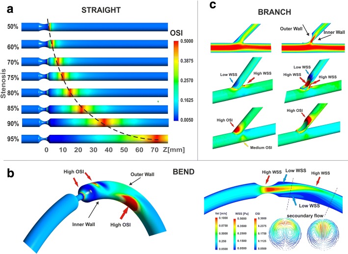
The results confirm our preliminary assumptions. There is significant variation in the coronary hemodynamic indices value caused by disturbed flow through stenosis in relation to variable flow conditions and geometry of vessels.

CONCLUSION

Variations of selected hemodynamic indexes induced by change of flow rate, heart rate and vessel geometry, obtained during a non-invasive study, may assist in evaluating the risk of stenosis progression and in carrying out the assessment of the hemodynamic significance of coronary stenosis. However, for a more accurate assessment of the variability of indices and coronary stenosis severity both local (near the narrowing) and global (in side branches) studies should be used.

摘要

背景

冠状动脉狭窄通常由动脉粥样硬化引起。通过比较血流紊乱所诱导的血流动力学效应,可评估特定患者冠状动脉狭窄的血流动力学意义及其进展风险。本研究展示了冠状动脉狭窄程度和血流条件变化如何影响振荡剪切指数、停留时间指数、压降系数和血流储备分数。我们假设,利用计算流体动力学评估的血流动力学指数相对于血流条件和血管几何形状的变化,可能是对多层计算机断层扫描检测到的冠状动脉狭窄进行无创评估的一个额外因素。

方法

使用血管基本形状(如直管段、弯曲段和分叉处)的局部参数化模型以及左冠状动脉的整体患者特异性模型,对虚拟重建的狭窄血管中的血流进行数值模拟。对无狭窄血管以及狭窄程度为10%至95%的血管进行计算。流速在20至1000毫升/分钟范围内变化,心率频率在30至210次/分钟范围内变化。基于有限元方法并经速度剖面实验测量验证的计算流体动力学,用于分析冠状动脉中的血流。

结果

结果证实了我们的初步假设。与血流条件和血管几何形状变化相关的、通过狭窄处的紊乱血流导致冠状动脉血流动力学指数值存在显著差异。

结论

在无创研究中获得的、由流速、心率和血管几何形状变化所诱导的选定血流动力学指标的变化,可能有助于评估狭窄进展风险以及对冠状动脉狭窄的血流动力学意义进行评估。然而,为了更准确地评估指标变异性和冠状动脉狭窄严重程度,应同时采用局部(狭窄附近)和整体(侧支内)研究。

https://cdn.ncbi.nlm.nih.gov/pmc/blobs/90f2/6025704/9e84462f5944/12872_2018_865_Fig12_HTML.jpg
https://cdn.ncbi.nlm.nih.gov/pmc/blobs/90f2/6025704/18aec57d2459/12872_2018_865_Fig1_HTML.jpg
https://cdn.ncbi.nlm.nih.gov/pmc/blobs/90f2/6025704/427aa757fa5a/12872_2018_865_Fig2_HTML.jpg
https://cdn.ncbi.nlm.nih.gov/pmc/blobs/90f2/6025704/9ea71e50f686/12872_2018_865_Fig3_HTML.jpg
https://cdn.ncbi.nlm.nih.gov/pmc/blobs/90f2/6025704/072b2ebb6fd5/12872_2018_865_Fig4_HTML.jpg
https://cdn.ncbi.nlm.nih.gov/pmc/blobs/90f2/6025704/ba7aea09fb26/12872_2018_865_Fig5_HTML.jpg
https://cdn.ncbi.nlm.nih.gov/pmc/blobs/90f2/6025704/98d12596e2d9/12872_2018_865_Fig6_HTML.jpg
https://cdn.ncbi.nlm.nih.gov/pmc/blobs/90f2/6025704/9ad54b2bd22c/12872_2018_865_Fig7_HTML.jpg
https://cdn.ncbi.nlm.nih.gov/pmc/blobs/90f2/6025704/a67a3ec1ff21/12872_2018_865_Fig8_HTML.jpg
https://cdn.ncbi.nlm.nih.gov/pmc/blobs/90f2/6025704/5157995786af/12872_2018_865_Fig9_HTML.jpg
https://cdn.ncbi.nlm.nih.gov/pmc/blobs/90f2/6025704/16cf0038b778/12872_2018_865_Fig10_HTML.jpg
https://cdn.ncbi.nlm.nih.gov/pmc/blobs/90f2/6025704/48181b623384/12872_2018_865_Fig11_HTML.jpg
https://cdn.ncbi.nlm.nih.gov/pmc/blobs/90f2/6025704/9e84462f5944/12872_2018_865_Fig12_HTML.jpg

相似文献

1
Numerical analysis of the impact of flow rate, heart rate, vessel geometry, and degree of stenosis on coronary hemodynamic indices.流速、心率、血管几何形状和狭窄程度对冠状动脉血流动力学指标影响的数值分析。
BMC Cardiovasc Disord. 2018 Jun 28;18(1):132. doi: 10.1186/s12872-018-0865-6.
2
Assessment of endothelial shear stress in patients with mild or intermediate coronary stenoses using coronary computed tomography angiography: comparison with invasive coronary angiography.使用冠状动脉计算机断层扫描血管造影术评估轻度或中度冠状动脉狭窄患者的内皮剪切应力:与有创冠状动脉造影术的比较。
Int J Cardiovasc Imaging. 2017 Jul;33(7):1101-1110. doi: 10.1007/s10554-016-1003-0. Epub 2016 Oct 31.
3
Fractional flow reserve-based 4D hemodynamic simulation of time-resolved blood flow in left anterior descending coronary artery.基于血流储备分数的左前降支冠状动脉时间分辨血流的4D血流动力学模拟
Clin Biomech (Bristol). 2019 Dec;70:164-169. doi: 10.1016/j.clinbiomech.2019.09.003. Epub 2019 Sep 10.
4
CT-based total vessel plaque analyses improves prediction of hemodynamic significance lesions as assessed by fractional flow reserve in patients with stable angina pectoris.基于 CT 的全血管斑块分析可改善对稳定性心绞痛患者血流动力学意义病变的预测,这些病变通过血流储备分数进行评估。
J Cardiovasc Comput Tomogr. 2018 Jul-Aug;12(4):344-349. doi: 10.1016/j.jcct.2018.04.008. Epub 2018 May 8.
5
Noninvasive CT-Derived FFR Based on Structural and Fluid Analysis: A Comparison With Invasive FFR for Detection of Functionally Significant Stenosis.基于结构和流体分析的无创 CT 衍生 FFR:与有创 FFR 检测功能显著狭窄的比较。
JACC Cardiovasc Imaging. 2017 Jun;10(6):663-673. doi: 10.1016/j.jcmg.2016.07.005. Epub 2016 Oct 19.
6
The impact of image resolution on computation of fractional flow reserve: coronary computed tomography angiography versus 3-dimensional quantitative coronary angiography.图像分辨率对血流储备分数计算的影响:冠状动脉计算机断层扫描血管造影与三维定量冠状动脉造影的比较
Int J Cardiovasc Imaging. 2016 Mar;32(3):513-23. doi: 10.1007/s10554-015-0797-5. Epub 2015 Oct 27.
7
Coronary Artery Axial Plaque Stress and its Relationship With Lesion Geometry: Application of Computational Fluid Dynamics to Coronary CT Angiography.冠状动脉轴向斑块应力及其与病变几何形状的关系:计算流体动力学在冠状动脉 CT 血管造影中的应用。
JACC Cardiovasc Imaging. 2015 Oct;8(10):1156-1166. doi: 10.1016/j.jcmg.2015.04.024. Epub 2015 Sep 9.
8
The Comparative Method Based on Coronary Computed Tomography Angiography for Assessing the Hemodynamic Significance of Coronary Artery Stenosis.基于冠状动脉计算机断层血管造影的冠状动脉狭窄血流动力学意义评估的比较方法。
Cardiovasc Eng Technol. 2023 Jun;14(3):364-379. doi: 10.1007/s13239-023-00658-2. Epub 2023 Mar 3.
9
Patient-Specific Hemodynamics of New Coronary Artery Bypass Configurations.新型冠状动脉旁路移植构型的患者特异性血液动力学。
Cardiovasc Eng Technol. 2020 Dec;11(6):663-678. doi: 10.1007/s13239-020-00493-9. Epub 2020 Oct 13.
10
Diagnostic performance of quantitative coronary computed tomography angiography and quantitative coronary angiography to predict hemodynamic significance of intermediate-grade stenoses.定量冠状动脉计算机断层扫描血管造影术和定量冠状动脉造影术预测中度狭窄血流动力学意义的诊断性能。
Int J Cardiovasc Imaging. 2015 Dec;31(8):1651-61. doi: 10.1007/s10554-015-0748-1. Epub 2015 Sep 1.

引用本文的文献

1
Comparison of blood viscosity models in different degrees of carotid artery stenosis.不同程度颈动脉狭窄时血液黏度模型的比较
PeerJ. 2025 Apr 28;13:e19336. doi: 10.7717/peerj.19336. eCollection 2025.
2
Hemodynamic effects of stenosis with varying severity in different segments of the carotid artery using computational fluid dynamics.利用计算流体动力学研究颈动脉不同节段不同严重程度狭窄的血流动力学效应。
Sci Rep. 2025 Feb 10;15(1):4896. doi: 10.1038/s41598-025-89100-2.
3
Narrowings of the Deep Cerebral Perforating Arteries Ostia: Geometry, Structure, and Clinical Implications.

本文引用的文献

1
Noninvasive FFR Derived From Coronary CT Angiography: Management and Outcomes in the PROMISE Trial.基于冠状动脉 CT 血管造影的无创血流储备分数:PROMISE 试验的处理和结果。
JACC Cardiovasc Imaging. 2017 Nov;10(11):1350-1358. doi: 10.1016/j.jcmg.2016.11.024. Epub 2017 Apr 12.
2
Numerical analysis of the effect of turbulence transition on the hemodynamic parameters in human coronary arteries.湍流转变对人体冠状动脉血流动力学参数影响的数值分析。
Cardiovasc Diagn Ther. 2016 Jun;6(3):208-20. doi: 10.21037/cdt.2016.03.08.
3
Proximal LAD atherosclerosis: milking-like effect of the septal perforator branches.
大脑深部穿支动脉开口狭窄:几何学、结构及临床意义
J Stroke. 2025 Jan;27(1):52-64. doi: 10.5853/jos.2024.01655. Epub 2025 Jan 31.
4
Non-invasive fractional flow reserve estimation in coronary arteries using angiographic images.基于冠状动脉造影图像的无创性冠状动脉血流储备分数估算。
Sci Rep. 2024 Jul 8;14(1):15640. doi: 10.1038/s41598-024-65626-9.
5
The Stress Phase Angle Measurement Using Spectral Domain Optical Coherence Tomography.利用谱域光学相干断层扫描测量应激相位角。
Sensors (Basel). 2023 Sep 1;23(17):7597. doi: 10.3390/s23177597.
6
Time-resolved simulation of blood flow through left anterior descending coronary artery: effect of varying extent of stenosis on hemodynamics.左前降支冠状动脉血流的时变模拟:狭窄程度变化对血液动力学的影响。
BMC Cardiovasc Disord. 2023 Mar 27;23(1):156. doi: 10.1186/s12872-023-03190-2.
7
The Comparative Method Based on Coronary Computed Tomography Angiography for Assessing the Hemodynamic Significance of Coronary Artery Stenosis.基于冠状动脉计算机断层血管造影的冠状动脉狭窄血流动力学意义评估的比较方法。
Cardiovasc Eng Technol. 2023 Jun;14(3):364-379. doi: 10.1007/s13239-023-00658-2. Epub 2023 Mar 3.
8
Machine Learning Identification Framework of Hemodynamics of Blood Flow in Patient-Specific Coronary Arteries with Abnormality.基于机器学习的冠状动脉血流动力学异常患者的个体化识别框架。
J Cardiovasc Transl Res. 2023 Jun;16(3):722-737. doi: 10.1007/s12265-022-10339-5. Epub 2022 Nov 18.
9
Fractional Flow Reserve (FFR) Estimation from OCT-Based CFD Simulations: Role of Side Branches.基于光学相干断层扫描(OCT)的计算流体动力学(CFD)模拟对血流储备分数(FFR)的估计:侧支血管的作用
Appl Sci (Basel). 2022 Jun;12(11). doi: 10.3390/app12115573. Epub 2022 May 30.
10
Non-invasive diagnostics of blockage growth in the descending aorta-computational approach.降主动脉阻塞生长的无创性诊断——计算方法。
Med Biol Eng Comput. 2022 Nov;60(11):3265-3279. doi: 10.1007/s11517-022-02665-2. Epub 2022 Sep 27.
左前降支近端动脉粥样硬化:间隔穿支的“挤奶样”效应
Jpn J Radiol. 2015 Aug;33(8):523-4. doi: 10.1007/s11604-015-0439-9. Epub 2015 Jun 17.
4
Clinical Relevance of Coronary Fractional Flow Reserve: Art-of-state.冠状动脉血流储备分数的临床相关性:现状
Chin Med J (Engl). 2015 May 20;128(10):1399-406. doi: 10.4103/0366-6999.156805.
5
Heart disease and stroke statistics--2015 update: a report from the American Heart Association.《2015年心脏病和中风统计数据更新:美国心脏协会报告》
Circulation. 2015 Jan 27;131(4):e29-322. doi: 10.1161/CIR.0000000000000152. Epub 2014 Dec 17.
6
Biomechanical factors in atherosclerosis: mechanisms and clinical implications.动脉粥样硬化中的生物力学因素:机制与临床意义
Eur Heart J. 2014 Nov 14;35(43):3013-20, 3020a-3020d. doi: 10.1093/eurheartj/ehu353. Epub 2014 Sep 17.
7
Numerical investigation of blood flow in three-dimensional porcine left anterior descending artery with various stenoses.三维猪左前降支不同狭窄程度下的血流数值研究。
Comput Biol Med. 2014 Apr;47:130-8. doi: 10.1016/j.compbiomed.2014.01.001. Epub 2014 Feb 11.
8
Invasive and non-invasive fractional flow reserve index in validation of hemodynamic severity of intracoronary lesions.有创和无创血流储备分数指数在冠状动脉内病变血流动力学严重程度验证中的应用
Postepy Kardiol Interwencyjnej. 2013;9(2):160-9. doi: 10.5114/pwki.2013.35452. Epub 2013 Jun 17.
9
Computational fluid dynamics applied to cardiac computed tomography for noninvasive quantification of fractional flow reserve: scientific basis.计算流体动力学在心脏计算机断层扫描中的应用,用于无创定量分数流量储备:科学基础。
J Am Coll Cardiol. 2013 Jun 4;61(22):2233-41. doi: 10.1016/j.jacc.2012.11.083. Epub 2013 Apr 3.
10
Does low and oscillatory wall shear stress correlate spatially with early atherosclerosis? A systematic review.低壁切应力和振荡壁切应力是否与早期动脉粥样硬化具有空间相关性?一项系统综述。
Cardiovasc Res. 2013 Jul 15;99(2):242-50. doi: 10.1093/cvr/cvt044. Epub 2013 Mar 3.