Department of Radiology and the Research Institute of Radiology, University of Ulsan College of Medicine, Asan Medical Center, Seoul 05505, Korea.
Department of Radiology, Korea University Ansan Hospital, Korea University College of Medicine, Ansan 15355, Korea.
Korean J Radiol. 2018 Jul-Aug;19(4):792-802. doi: 10.3348/kjr.2018.19.4.792. Epub 2018 Jun 14.
To describe CT and clinical findings of pulmonary artery intimal sarcoma (PAIS) compared with those of pulmonary thromboembolism (PTE), to investigate MRI and positron emission tomography (PET)-CT findings of PAIS, and to evaluate the effect of delayed diagnosis of PAIS on survival outcomes.
Twenty-six patients with PAIS were retrospectively identified and matched for sex, with patients with PTE at a ratio of 1:2. CT and clinical findings of the two groups were compared using Student's test or chi-square test. The effect of delayed diagnosis on survival was investigated using Kaplan-Meier analysis.
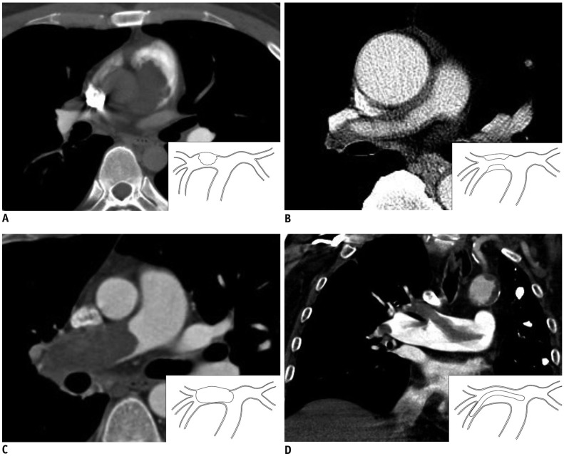
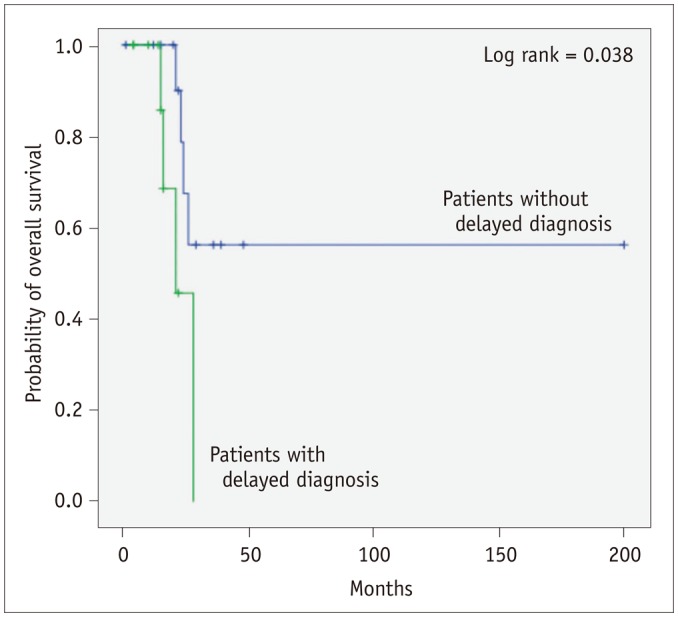
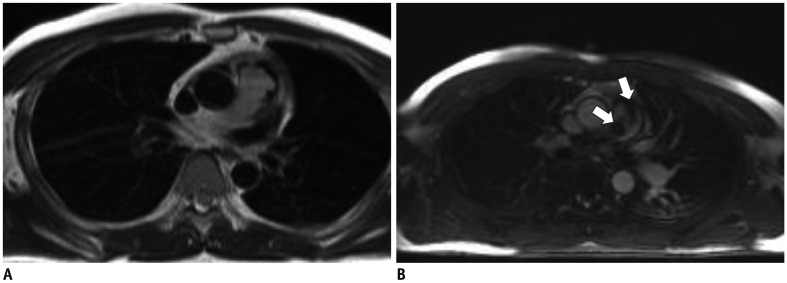
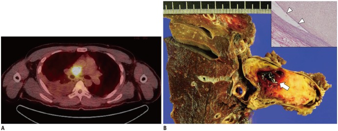
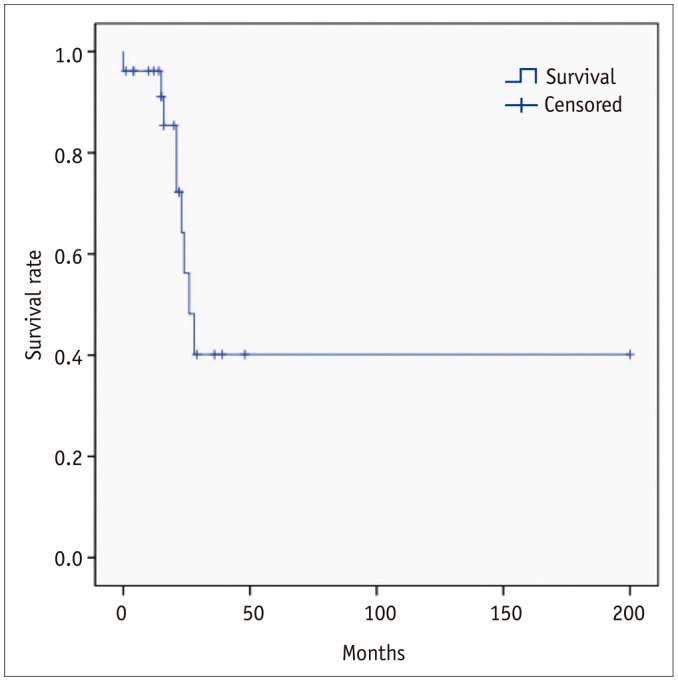
The most common tumor pattern in PAIS was tumoral impaction. Heterogeneous attenuation, wall eclipse signs, intratumoral vessels, acute interphase angles, single location, presence of lung ischemia, and central location were significantly more common in PAIS than in PTE (all < 0.01). Levels of D-dimers and brain natriuretic peptide were lower in PAIS than in PTE ( < 0.05). In three patients of PAIS, long inversion time sequence MRI showed intermingled dark signal intensity foci suggestive of intermingled thrombi. All nine patients who had undergone PET-CT displayed hypermetabolism. Diagnosis was delayed in 42.3% of the PAIS patients and those patients had a significantly shorter overall survival than patients whose diagnosis was not delayed ( < 0.05).
The characteristic CT and clinical findings of PAIS may help achieve early diagnosis of PAIS and make better survival outcomes of patients. MRI and PET-CT can be used as second-line imaging modalities and could help distinguish PAIS from PTE and to plan clinical management.
描述肺动脉内膜肉瘤(PAIS)的 CT 和临床特征,并与肺血栓栓塞症(PTE)进行比较,探讨 MRI 和正电子发射断层扫描(PET)-CT 在 PAIS 中的表现,并评估 PAIS 延迟诊断对生存结局的影响。
回顾性地确定了 26 例 PAIS 患者,并按性别与 PTE 患者进行了匹配,比例为 1:2。使用 Student's 检验或卡方检验比较两组的 CT 和临床特征。使用 Kaplan-Meier 分析研究延迟诊断对生存的影响。
PAIS 最常见的肿瘤模式是肿瘤嵌塞。与 PTE 相比,PAIS 中更常见的 CT 表现包括不均匀衰减、壁影征、瘤内血管、急性交界角、单发部位、存在肺缺血和中央部位(均 < 0.01)。PAIS 患者的 D-二聚体和脑钠肽水平低于 PTE 患者(均 < 0.05)。在 3 例 PAIS 患者中,长反转时间序列 MRI 显示混杂的暗信号强度灶,提示混杂血栓。9 例接受 PET-CT 检查的患者均显示高代谢。PAIS 患者中有 42.3%的诊断延迟,与诊断不延迟的患者相比,这些患者的总生存时间明显更短(< 0.05)。
PAIS 的特征性 CT 和临床特征有助于早期诊断 PAIS,并改善患者的生存结局。MRI 和 PET-CT 可作为二线成像方式,有助于区分 PAIS 和 PTE,并为临床管理提供依据。