Department of Physiology, Hokkaido University School of Medicine, Sapporo, Japan.
Laboratory of Sensorimotor Research, National Eye Institute, National Institutes of Health, Bethesda, United States.
Elife. 2018 Jul 2;7:e35676. doi: 10.7554/eLife.35676.
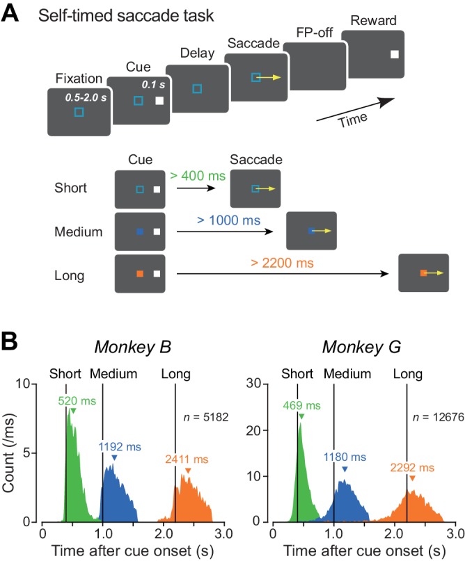
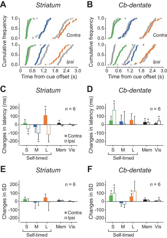
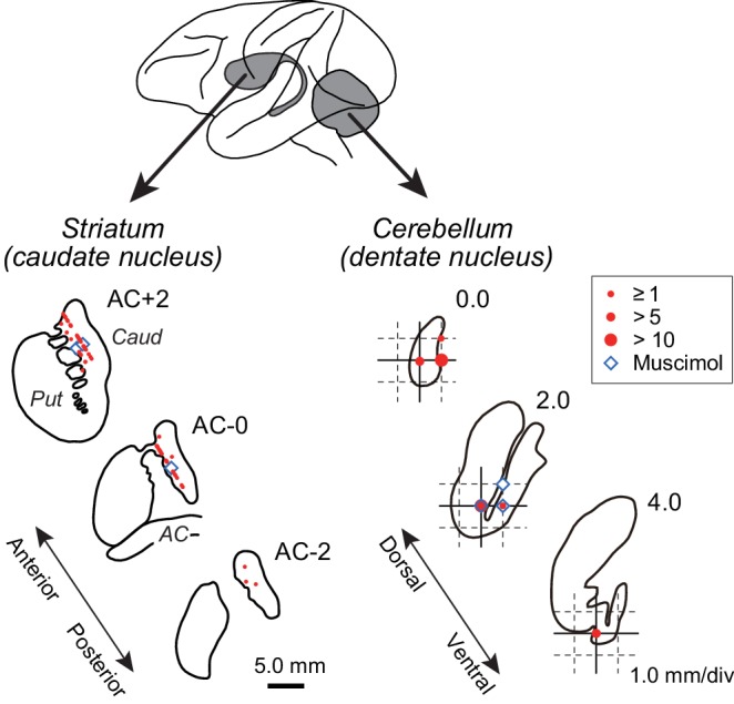
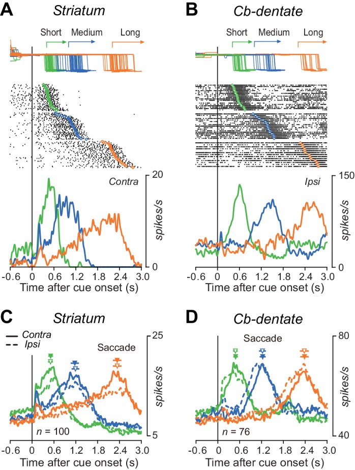
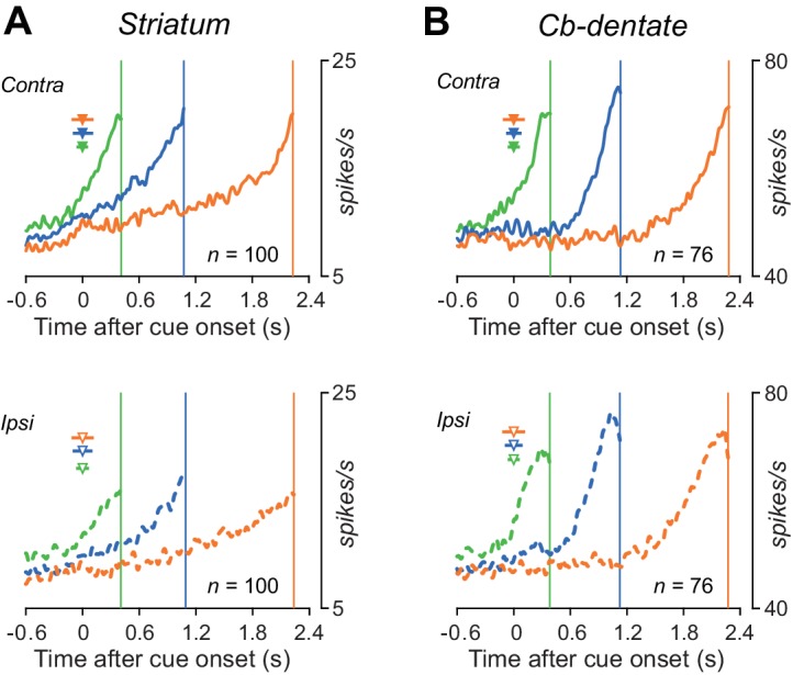
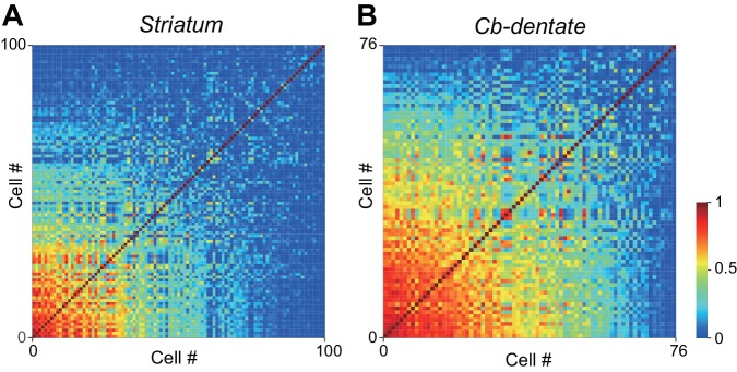
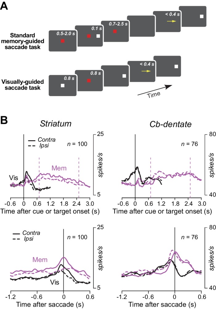
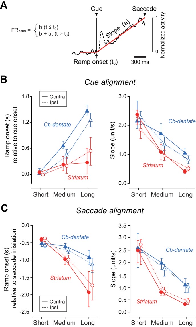
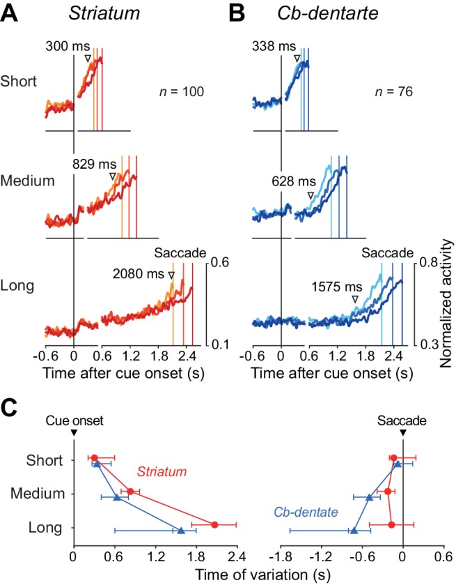
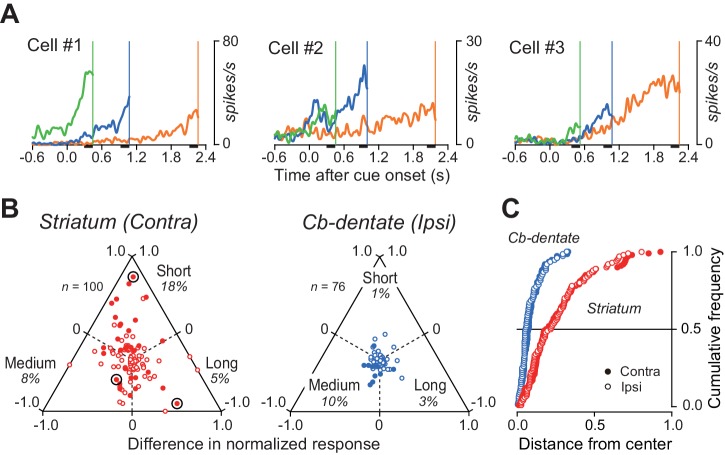
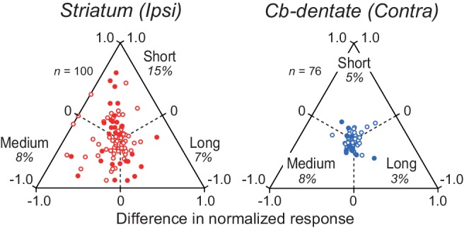
The ability to flexibly adjust movement timing is important for everyday life. Although the basal ganglia and cerebellum have been implicated in monitoring of supra- and sub-second intervals, respectively, the underlying neuronal mechanism remains unclear. Here, we show that in monkeys trained to generate a self-initiated saccade at instructed timing following a visual cue, neurons in the caudate nucleus kept track of passage of time throughout the delay period, while those in the cerebellar dentate nucleus were recruited only during the last part of the delay period. Conversely, neuronal correlates of trial-by-trial variation of self-timing emerged earlier in the cerebellum than the striatum. Local inactivation of respective recording sites confirmed the difference in their relative contributions to supra- and sub-second intervals. These results suggest that the basal ganglia may measure elapsed time relative to the intended interval, while the cerebellum might be responsible for the fine adjustment of self-timing.
灵活调整运动时间的能力对日常生活很重要。尽管基底神经节和小脑分别被认为与超秒和亚秒间隔的监测有关,但潜在的神经元机制仍不清楚。在这里,我们发现在猴子被训练以在视觉提示后按照指令时间自主产生扫视时,尾状核中的神经元在整个延迟期间都能记录时间的流逝,而齿状核中的神经元则仅在延迟期间的最后部分被募集。相反,自我定时的逐次试验变化的神经元相关性在小脑比纹状体更早出现。在各自的记录部位进行局部失活确认了它们对超秒和亚秒间隔的相对贡献的差异。这些结果表明,基底神经节可能会测量相对于预期间隔的经过时间,而小脑可能负责自我定时的精细调整。