Department of Radiology, the First Affiliated Hospital, Xi'an Jiaotong University Xi'an, #277, Yanta West Road, Xi'an, 710061, Shaanxi, China.
Department of Radiology, the Northwest Women and Children Hospital, #1616, Yanxiang Road, Xi'an, 710054, Shaanxi, China.
Cancer Imaging. 2018 Jul 3;18(1):23. doi: 10.1186/s40644-018-0156-6.
The invasion depth of endometrial cancer is one of the most important prognosis factors. The aim of the current study was to investigate the diagnostic value of the apparent diffusion coefficient (ADC) of the peritumoral zone for assessing the infiltration depth of endometrial cancer.
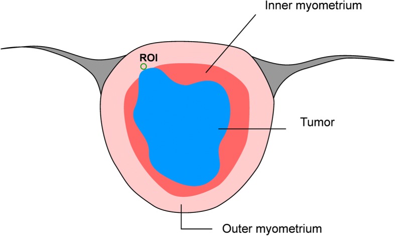
An institutional review board approved this prospective study, and all study participants provided informed consent. A total of 58 patients (mean age 54 ± 8.3 years, range 34-69 years) with endometrial cancer were prospectively enrolled. Two radiologists assessed all preoperative magnetic resonance images with T1, T2, and diffusion-weighted imaging, and determined the location of the deepest invasion of the tumor. The peritumoral zone was defined as a 5-mm-thick zone surrounding and adjacent to the cancerous endometrium. The mean ADC (ADCm) values of the tumor and the peritumoral zone were measured. Sensitivity, specificity, positive and negative predictive values, and the area under the receiver operating characteristic curve (Az) were calculated for visual inspection, and an ADC cutoff value for the peri-endometrial zone was determined for predicting the myometrial invasion depth.
The ADCm values of tumors and peritumoral zones were 0.83 × 10 mm/sec and 1.06 × 10 mm/sec, respectively. There was no significant difference between the ADCm values of the tumors in the superficial and deep myometrial invasion groups (P > 0.05). However, the ADCm value at the peritumoral zone in the deep myometrial invasion group (1.23 × 10 mm/sec) significantly differed from that in the superficial myometrial invasion group (0.99 × 10 mm/sec) (p = 0.005). In assessments of deep myometrial invasion, the sensitivity, specificity, negative predictive value, and positive predictive value were 0.58, 0.93, 0.84, and 0.77, respectively, for the ADCm cutoff value of the peritumoral zone, and 0.71, 0.80, 0.87, and 0.60. respectively, for visual inspection. The accuracy of myometrial invasion depth assessment using the ADCm cutoff value and visual inspection were 83 and 78%, respectively. The Az for both was 0.76.
ADCm at the peritumoral zone can predict deep myometrial invasion of endometrial cancer. This value can therefore enhance confidence in preoperative endometrial cancer evaluation, and when tailoring surgical approaches.
子宫内膜癌的浸润深度是最重要的预后因素之一。本研究旨在探讨肿瘤周围区表观扩散系数(ADC)评估子宫内膜癌浸润深度的诊断价值。
本研究经机构审查委员会批准,所有研究参与者均提供了知情同意书。前瞻性纳入 58 例子宫内膜癌患者(平均年龄 54±8.3 岁,范围 34-69 岁)。两名放射科医生评估了所有术前 T1、T2 和弥散加权成像的磁共振图像,并确定了肿瘤最深浸润的位置。肿瘤周围区定义为环绕和毗邻癌性子宫内膜的 5-mm 厚的区域。测量肿瘤和肿瘤周围区的平均 ADC(ADCm)值。通过视觉检查计算灵敏度、特异性、阳性和阴性预测值以及受试者工作特征曲线下面积(Az),并确定肿瘤周围区 ADC 截断值以预测肌层浸润深度。
肿瘤和肿瘤周围区的 ADCm 值分别为 0.83×10mm/sec 和 1.06×10mm/sec。在浅肌层浸润组和深肌层浸润组之间,肿瘤的 ADCm 值没有显著差异(P>0.05)。然而,深肌层浸润组肿瘤周围区的 ADCm 值(1.23×10mm/sec)明显不同于浅肌层浸润组(0.99×10mm/sec)(p=0.005)。在评估深肌层浸润时,肿瘤周围区 ADCm 截断值的灵敏度、特异性、阴性预测值和阳性预测值分别为 0.58、0.93、0.84 和 0.77,而视觉检查分别为 0.71、0.80、0.87 和 0.60。使用 ADCm 截断值和视觉检查评估肌层浸润深度的准确性分别为 83%和 78%。两者的 Az 均为 0.76。
肿瘤周围区的 ADCm 可预测子宫内膜癌的深肌层浸润。因此,该值可以增强术前对子宫内膜癌的评估信心,并有助于制定手术方法。