Nakai Nobuhiro, Takumi Toru, Nakai Junichi, Sato Masaaki
RIKEN Center for Brain Science, Wako, Japan.
RIKEN Center for Advanced Intelligence Project, Tokyo, Japan.
Front Neurosci. 2018 Jun 19;12:412. doi: 10.3389/fnins.2018.00412. eCollection 2018.
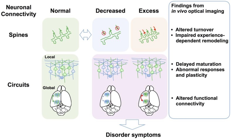
optical imaging is a powerful tool for revealing brain structure and function at both the circuit and cellular levels. Here, we provide a systematic review of findings obtained from imaging studies of mouse models of neurodevelopmental disorders, including the monogenic disorders fragile X syndrome, Rett syndrome, and Angelman syndrome, which are caused by genetic abnormalities of , and , as well as disorders caused by copy number variations (15q11-13 duplication and 22q11.2 deletion) and BTBR mice as an inbred strain model of autism spectrum disorder (ASD). Most studies visualize the structural and functional responsiveness of cerebral cortical neurons to sensory stimuli and the developmental and experience-dependent changes in these responses as a model of brain functions affected by these disorders. The optical imaging techniques include two-photon microscopy of fluorescently labeled dendritic spines or neurons loaded with fluorescent calcium indicators and macroscopic imaging of cortical activity using calcium indicators, voltage-sensitive dyes or intrinsic optical signals. Studies have revealed alterations in the density, stability, and turnover of dendritic spines, aberrant cortical sensory responses, impaired inhibitory function, and concomitant failure of circuit maturation as common causes for neurological deficits. Mechanistic hypotheses derived from imaging also provide new directions for therapeutic interventions. For instance, it was recently demonstrated that early postnatal administration of a selective serotonin reuptake inhibitor (SSRI) restores impaired cortical inhibitory function and ameliorates the aberrant social behaviors in a mouse model of ASD. We discuss the potential use of SSRIs for treating ASDs in light of these findings.
光学成像是在回路和细胞水平揭示脑结构与功能的强大工具。在此,我们对神经发育障碍小鼠模型成像研究的结果进行系统综述,这些模型包括由、和的基因异常引起的单基因疾病脆性X综合征、雷特综合征和天使综合征,以及由拷贝数变异(15q11 - 13重复和22q11.2缺失)导致的疾病,还有作为自闭症谱系障碍(ASD)近交系模型的BTBR小鼠。大多数研究将大脑皮质神经元对感觉刺激的结构和功能反应性以及这些反应中依赖发育和经验的变化视为受这些疾病影响的脑功能模型进行可视化。光学成像技术包括对用荧光钙指示剂标记的树突棘或神经元进行双光子显微镜成像,以及使用钙指示剂、电压敏感染料或内在光学信号对皮质活动进行宏观成像。研究揭示了树突棘密度、稳定性和更新的改变、异常的皮质感觉反应、抑制功能受损以及回路成熟伴随失败是神经功能缺陷的常见原因。从成像得出的机制假说也为治疗干预提供了新方向。例如,最近证明在出生后早期给予选择性5-羟色胺再摄取抑制剂(SSRI)可恢复ASD小鼠模型中受损的皮质抑制功能并改善异常的社会行为。鉴于这些发现,我们讨论了SSRI治疗ASD的潜在用途。