Frangiamore Salvatore J, Morris Elizabeth R, Scibetta Alex C, Chahla Jorge, Moatshe Gilbert, Civitarese David, Provencher Matthew T, Hackett Thomas R, Schickendantz Mark S, Huard Johnny, LaPrade Robert F
The Steadman Clinic, Vail, Colorado, USA.
Steadman Philippon Research Institute, Vail, Colorado, USA.
Orthop J Sports Med. 2018 Jun 12;6(6):2325967118777825. doi: 10.1177/2325967118777825. eCollection 2018 Jun.
Vascular-derived progenitor and endothelial cell populations (CD31, CD34, CD146) are capable of multipotent differentiation at the site of injured ligamentous tissue to aid in the intrinsic healing response. Proximal ulnar collateral ligament (UCL) tears have been reported to have better healing capability when compared with distal UCL tears.
To compare the vascular composition of the proximal and distal insertions of the anterior bundle of the UCL of the elbow via known markers of endothelial and vascular-derived progenitor cells (CD31, CD34, CD146).
Descriptive laboratory study.
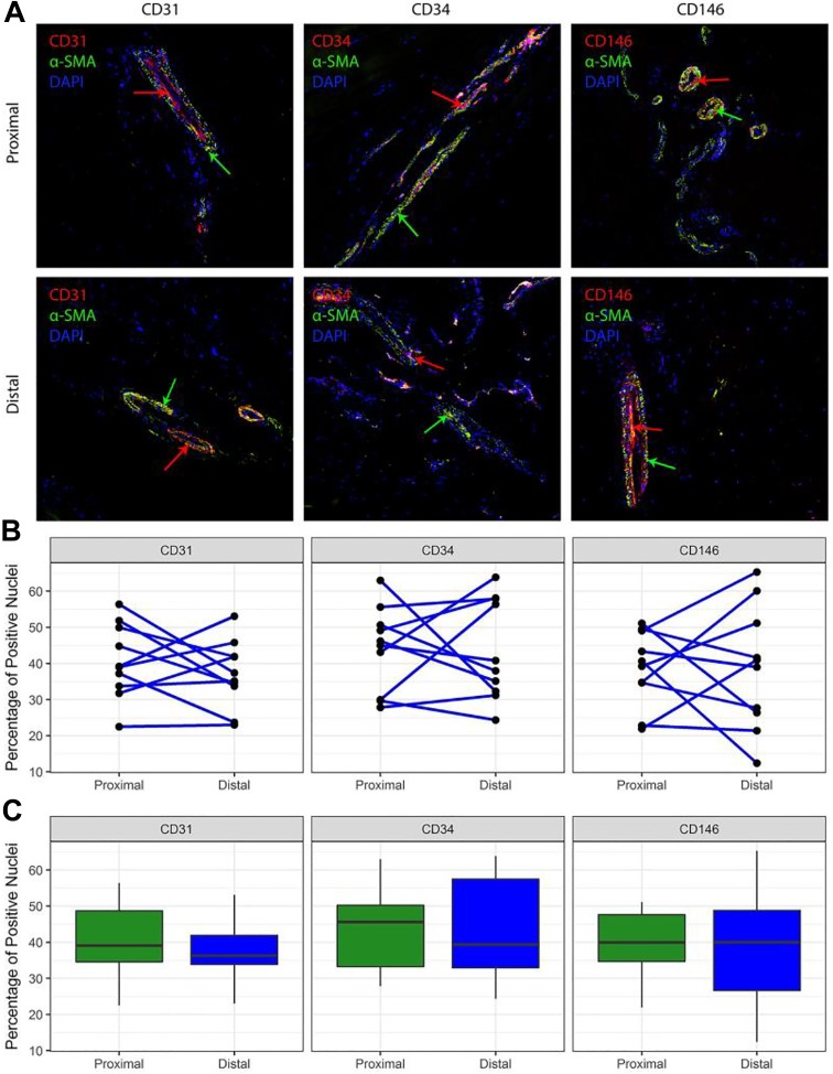
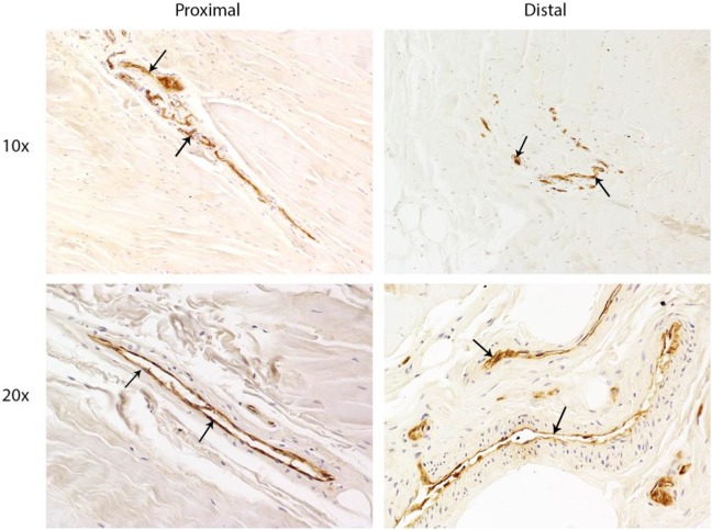
UCLs were harvested from 10 nonpaired fresh-frozen human cadaveric elbows and transected into proximal and distal portions. Endothelial and vascular-derived progenitor cell densities were assessed with 4 staining groups: CD31 (immunohistochemistry) and CD31/α-smooth muscle actin (α-SMA), CD34/α-SMA, and CD146/α-SMA (immunofluorescence). CD31 immunohistochemistry identified endothelial progenitor cells in the UCL. Later staining of the same slides with α-SMA demonstrated the relationship of progenitor cells to the surrounding vasculature. Fluorescent staining was quantified by calculating the proportion of positively stained nuclei versus the total number of nuclei in the proximal and distal UCL.
CD31+ cells were present in the proximal and distal sections of all 10 UCLs. Fluorescent staining revealed no significant differences in the ratio of CD31 to total nuclei between the distal (median, 36% [range, 23%-53%]) and proximal UCL (39% [22%-56%]) ( = .432, Wilcoxon signed-rank test). Similarly, no differences were seen between CD34 distal (39% [24%-64%]) and proximal regions (46% [28%-63%]) ( = .846, Wilcoxon signed-rank test) or CD146 distal (40% [12%-65%]) and proximal regions (40% [22%-51%]) ( ≥ .999, Wilcoxon signed-rank test).
Analysis of UCL tissues demonstrated equal distributions of vascular endothelial and vascular-derived progenitor cell markers throughout the proximal and distal UCL. Unlike that of the medial collateral ligament of the knee, the microvascular composition of the proximal and distal UCL insertions was not different, suggesting a well-vascularized ligament throughout its course.
These findings investigate one of the possible contributors to UCL healing after injury, which may provide insight into operative and nonoperative management of UCL injuries in the future. This study also indicates that reasons other than differences in progenitor cell density alone may explain the clinical healing differences seen between proximal and distal UCL tears. A better understanding of the microvascular environment and associated blood supply is warranted to understand the healing capability of the UCL.
血管源性祖细胞和内皮细胞群体(CD31、CD34、CD146)能够在损伤的韧带组织部位进行多能分化,以辅助内在愈合反应。据报道,与尺侧副韧带(UCL)远端撕裂相比,近端撕裂具有更好的愈合能力。
通过内皮细胞和血管源性祖细胞的已知标志物(CD31、CD34、CD146)比较肘部UCL前束近端和远端附着点的血管组成。
描述性实验室研究。
从10具非配对的新鲜冷冻人体尸体肘部获取UCL,并将其横断为近端和远端部分。用4个染色组评估内皮细胞和血管源性祖细胞密度:CD31(免疫组织化学)以及CD31/α-平滑肌肌动蛋白(α-SMA)、CD34/α-SMA和CD146/α-SMA(免疫荧光)。CD31免疫组织化学鉴定UCL中的内皮祖细胞。随后用α-SMA对同一张玻片进行染色,以显示祖细胞与周围脉管系统的关系。通过计算近端和远端UCL中阳性染色细胞核占总细胞核数的比例来对荧光染色进行定量。
所有10条UCL的近端和远端均存在CD31+细胞。荧光染色显示,远端UCL(中位数,36%[范围,23%-53%])和近端UCL(39%[22%-56%])之间CD31与总细胞核的比例无显著差异(P = 0.432,Wilcoxon符号秩和检验)。同样,CD34在远端(39%[24%-64%])和近端区域(46%[28%-63%])之间(P = 0.846,Wilcoxon符号秩和检验)或CD146在远端(40%[12%-65%])和近端区域(40%[22%-51%])之间(P≥0.999,Wilcoxon符号秩和检验)也无差异。
UCL组织分析表明,血管内皮细胞和血管源性祖细胞标志物在UCL近端和远端分布均匀。与膝关节内侧副韧带不同,UCL近端和远端附着点的微血管组成并无差异,这表明整个韧带的血管化良好。
这些发现探究了损伤后UCL愈合的可能影响因素之一,这可能为未来UCL损伤的手术和非手术治疗提供见解。本研究还表明,除了祖细胞密度差异之外的其他原因可能解释了UCL近端和远端撕裂在临床愈合上的差异。有必要更好地了解微血管环境及相关血供,以了解UCL的愈合能力。