Lee Seongpung, Kim Jun Young, Hong Jaesung, Baek Seung Hoon, Kim Shin Yoon
Department of Robotics Engineering, Daegu Gyeongbuk Institute of Science and Technology, Daegu, Korea.
Department of Orthopaedic Surgery, Daegu Catholic University College of Medicine, Daegu, Korea.
Yonsei Med J. 2018 Aug;59(6):769-780. doi: 10.3349/ymj.2018.59.6.769.
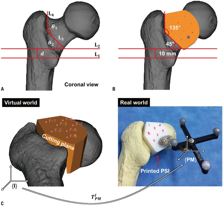
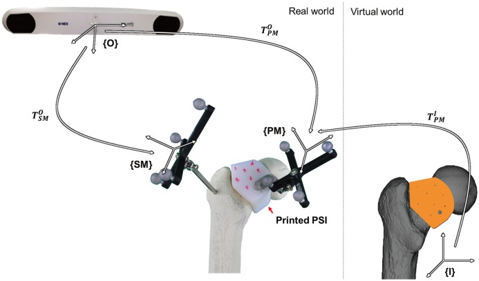
The intraoperative version of the femoral component is usually determined by visual appraisal of the stem position relative to the distal femoral condylar axis. However, several studies have suggested that a surgeon's visual assessment of the stem position has a high probability of misinterpretation. We developed a computed tomography (CT)-based navigation system with a patient-specific instrument (PSI) capable of three-dimensional (3D) printing and investigated its accuracy and consistency in comparison to the conventional technique of visual assessment of the stem position.
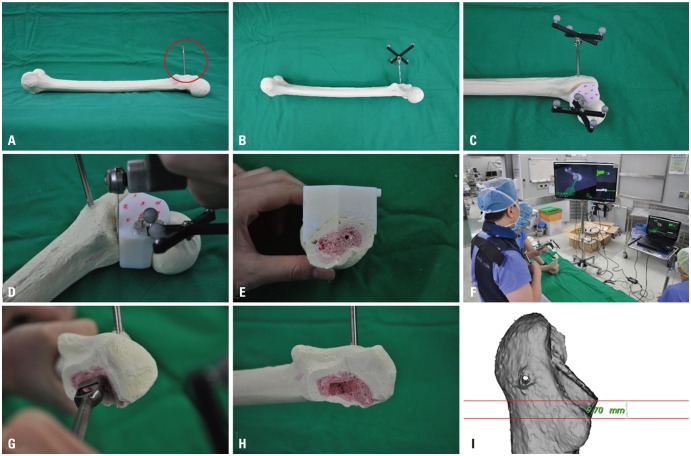
A CT scan of a femur sawbone model was performed, and pre-experimental planning was completed. We conducted 30 femoral neck osteotomies using the conventional technique and another 30 femoral neck osteotomies using the proposed technique. The femoral medullary canals were identified in both groups using a box chisel.
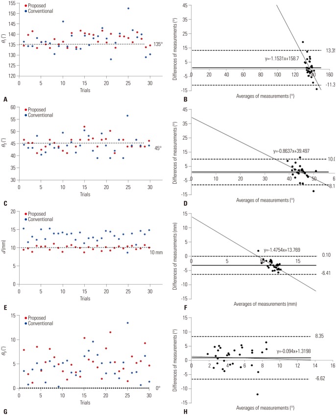
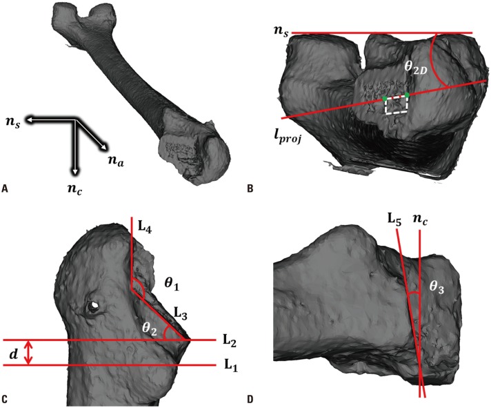
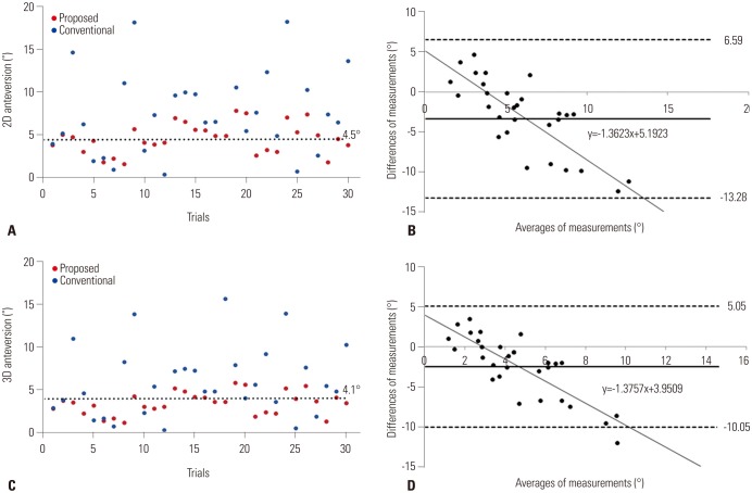
For the absolute deviation between the measured and planned values, the mean two-dimensional anteversions of the proposed and conventional techniques were 1.41° and 4.78°, while their mean 3D anteversions were 1.15° and 3.31°. The mean θ₁, θ₂, θ₃, and d, all of which are parameters for evaluating femoral neck osteotomy, were 2.93°, 1.96°, 5.29°, and 0.48 mm for the proposed technique and 4.26°, 3.17°, 4.43°, and 3.15 mm for the conventional technique, respectively.
The CT-based navigation system with PSI was more accurate and consistent than the conventional technique for assessment of stem position. Therefore, it can be used to reduce the frequency of incorrect assessments of the stem position among surgeons and to help with accurate determination of stem anteversion.
股骨部件的术中版本通常通过视觉评估柄相对于股骨远端髁轴的位置来确定。然而,多项研究表明,外科医生对柄位置的视觉评估很可能出现错误解读。我们开发了一种基于计算机断层扫描(CT)的导航系统,该系统配备了能够进行三维(3D)打印的患者特异性器械(PSI),并与传统的柄位置视觉评估技术相比,研究了其准确性和一致性。
对股骨锯骨模型进行CT扫描,并完成术前规划。我们使用传统技术进行了30例股骨颈截骨术,使用所提出的技术又进行了30例股骨颈截骨术。两组均使用盒凿识别股骨髓腔。
对于测量值与计划值之间的绝对偏差,所提出的技术和传统技术的平均二维前倾角分别为1.41°和4.78°,而它们的平均3D前倾角分别为1.15°和3.31°。用于评估股骨颈截骨术的所有参数,即平均θ₁、θ₂、θ₃和d,所提出的技术分别为2.93°、1.96°、5.29°和0.48mm,传统技术分别为4.26°、3.17°、4.43°和3.15mm。
带有PSI的基于CT的导航系统在评估柄位置方面比传统技术更准确、更一致。因此,它可用于减少外科医生对柄位置错误评估的频率,并有助于准确确定柄的前倾角。