Institute of Cardiovascular Sciences, University of Manchester, Manchester, UK.
Environment and Life Sciences, University of Salford, Salford, UK.
Br J Pharmacol. 2018 Sep;175(18):3685-3698. doi: 10.1111/bph.14433. Epub 2018 Aug 10.
In response to noradrenaline, healthy perivascular adipose tissue (PVAT) exerts an anticontractile effect on adjacent small arterial tissue. Organ bath solution transfer experiments have demonstrated the release of PVAT-derived relaxing factors that mediate this function. The present studies were designed to investigate the mechanism responsible for the noradrenaline-induced PVAT anticontractile effect.
In vitro rat small arterial contractile function was assessed using wire myography in the presence and absence of PVAT and the effects of sympathomimetic stimulation on the PVAT environment explored using Western blotting and assays of organ bath buffer.
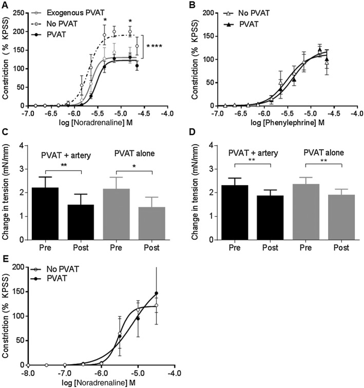
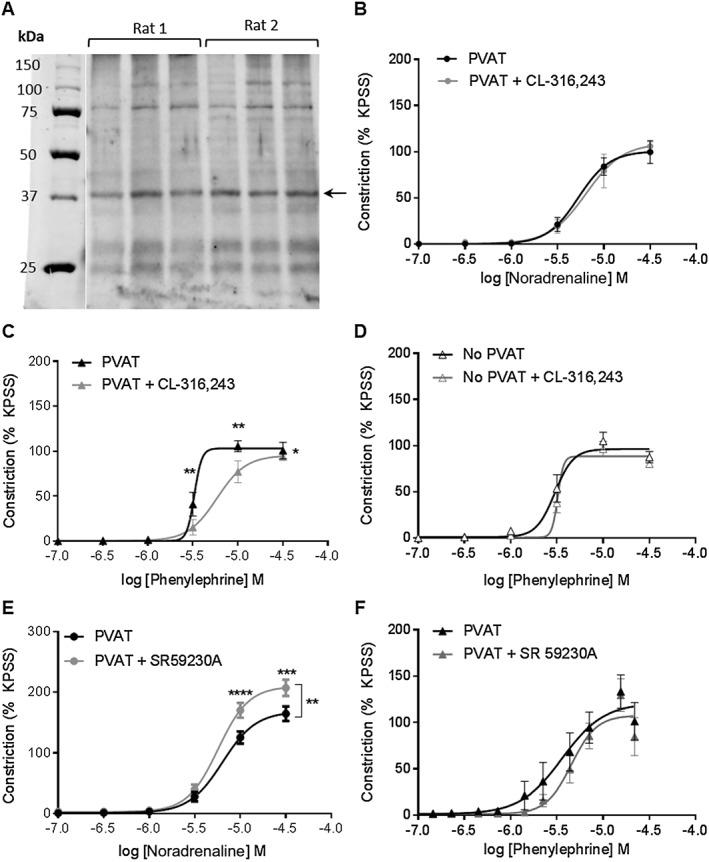
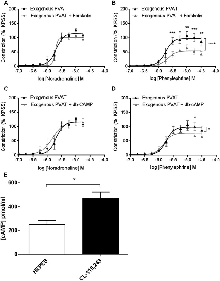
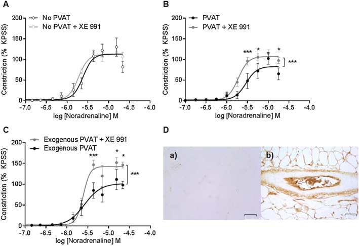
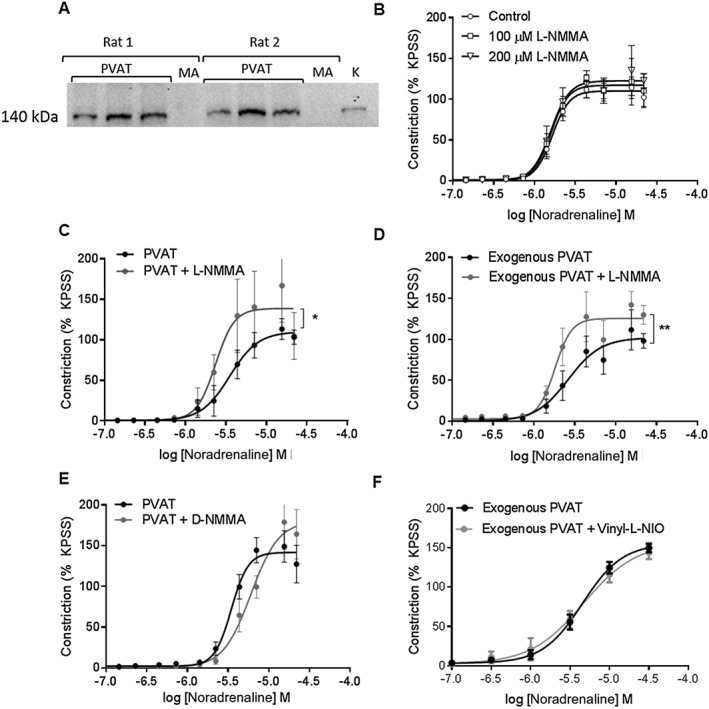
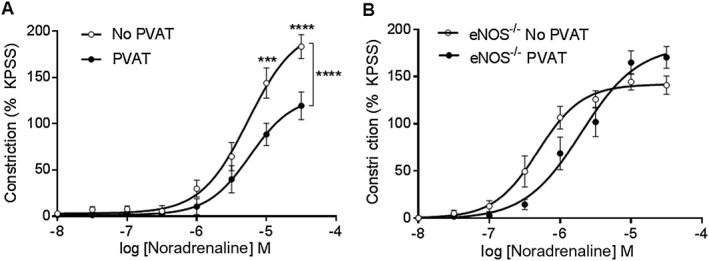
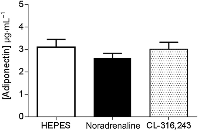
PVAT elicited an anticontractile effect in response to noradrenaline but not phenylephrine stimulation. In arteries surrounded by intact PVAT, the β -adrenoceptor agonist, CL-316243, reduced the vasoconstrictor effect of phenylephrine but not noradrenaline. K 7 channel inhibition using XE 991 reversed the noradrenaline-induced anticontractile effect in exogenously applied PVAT studies. Adrenergic stimulation of PVAT with noradrenaline and CL-316243, but not phenylephrine, was associated with increased adipocyte-derived NO production, and the contractile response to noradrenaline was augmented following incubation of exogenous PVAT with L-NMMA. PVAT from eNOS mice had no anticontractile effect. Assays of adipocyte cAMP demonstrated an increase with noradrenaline stimulation implicating Gα signalling in this process.
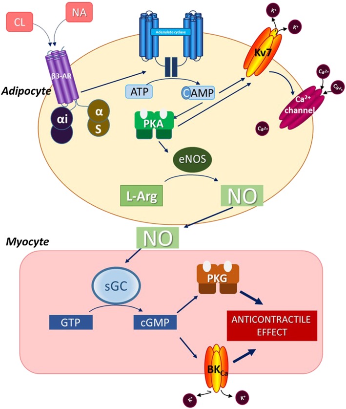
We have shown that adipocyte-located β -adrenoceptor stimulation leads to activation of Gα signalling pathways with increased cAMP and the release of adipocyte-derived NO. This process is dependent upon K 7 channel function. We conclude that adipocyte-derived NO plays a central role in anticontractile activity when rodent PVAT is stimulated by noradrenaline.
健康的血管周围脂肪组织(PVAT)在受到去甲肾上腺素刺激时会对邻近的小动脉组织产生抗收缩作用。器官浴液转移实验已经证明了 PVAT 衍生的舒张因子的释放,这些因子介导了这一功能。本研究旨在探讨负责去甲肾上腺素诱导的 PVAT 抗收缩作用的机制。
在存在和不存在 PVAT 的情况下,使用wire myography 评估体外大鼠小动脉收缩功能,并使用 Western blot 和器官浴液缓冲液测定研究交感神经刺激对 PVAT 环境的影响。
PVAT 对去甲肾上腺素刺激产生抗收缩作用,但对苯肾上腺素刺激无反应。在完整的 PVAT 包围的动脉中,β-肾上腺素受体激动剂 CL-316243 降低了苯肾上腺素的血管收缩作用,但不降低去甲肾上腺素的血管收缩作用。使用 XE 991 抑制 K 7 通道逆转了外源性应用 PVAT 研究中去甲肾上腺素诱导的抗收缩作用。用去甲肾上腺素和 CL-316243 刺激 PVAT 导致脂肪细胞衍生的 NO 产生增加,而用 L-NMMA 孵育外源性 PVAT 后,对去甲肾上腺素的收缩反应增强。来自 eNOS 小鼠的 PVAT 没有抗收缩作用。测定脂肪细胞 cAMP 表明,刺激去甲肾上腺素后 cAMP 增加,表明 Gα 信号参与了这一过程。
我们已经表明,脂肪细胞定位的β-肾上腺素受体刺激导致 Gα 信号通路的激活,增加 cAMP 和脂肪细胞衍生的 NO 的释放。这个过程依赖于 K 7 通道的功能。我们得出结论,当去甲肾上腺素刺激啮齿动物 PVAT 时,脂肪细胞衍生的 NO 在抗收缩活性中起核心作用。