Veronesi Bruno Azevedo, Rodrigues Marcelo Bordalo, Sambuy Marina Tommasini Carrara DE, Macedo Rodrigo Sousa, Cho Álvaro Baik, Rezende Marcelo Rosa DE
. Hand and Microsurgery Group, Instituto de Ortopedia e Traumatologia, Hospital das Clinicas HCFMUSP, Faculdade de Medicina, Universidade de São Paulo, São Paulo, SP, Brazil.
. Radiology Department, Instituto de Ortopedia e Traumatologia, Hospital das Clinicas HCFMUSP, Faculdade de Medicina, Universidade de São Paulo, São Paulo, SP, Brazil.
Acta Ortop Bras. 2018 Mar-Apr;26(2):131-134. doi: 10.1590/1413-785220182602187223.
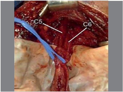
To compare magnetic resonance imaging and intraoperative findings in patients diagnosed with traumatic injury to the brachial plexus.
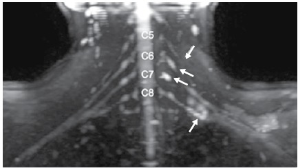
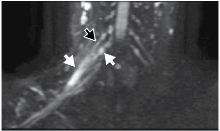
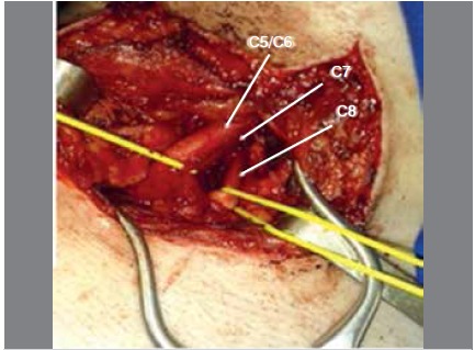
Patients with a diagnosis of traumatic injury to the brachial plexus admitted to the hand and microsurgery outpatient consult of the Hospital das Clínicas at the University of São Paulo were selected during December 2016. A total of three adult patients with up to six months of injury who underwent surgical treatment were included in the study. A diffusion-weighted sequence magnetic resonance protocol and fluid-sensitive volumetric reformatting sequence were applied. The magnetic resonance results were compared with the diagnoses obtained from the injuries observed during the surgery. The study was double-blind (surgeon and radiologist).
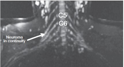
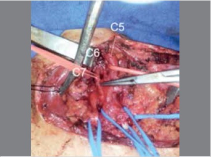
A descriptive correlation was found between the magnetic resonance imaging results and the diagnostic findings from the surgeries, for both pre- and post-ganglionic injuries.
Magnetic resonance imaging has shown to be a promising diagnostic method in preoperative assessment of brachial plexus lesions; it is less invasive than other common methods, showing not only avulsion lesions but also localized postganglionic lesions in the supra- and infraclavicular region. Level of Evidence III; Diagnostic studies - Investigating a diagnostic test.
比较诊断为臂丛神经创伤性损伤患者的磁共振成像结果与术中发现。
选取2016年12月期间在圣保罗大学临床医院手部和显微外科门诊咨询就诊、诊断为臂丛神经创伤性损伤的患者。共有3例受伤时间长达6个月且接受手术治疗的成年患者纳入研究。应用扩散加权序列磁共振成像方案和液体敏感容积重建成像序列。将磁共振成像结果与手术中观察到的损伤诊断结果进行比较。该研究为双盲研究(外科医生和放射科医生)。
对于节前和节后损伤,磁共振成像结果与手术诊断结果之间均发现有描述性相关性。
磁共振成像已被证明是臂丛神经损伤术前评估中有前景的诊断方法;它比其他常用方法侵入性小,不仅能显示撕脱伤,还能显示锁骨上和锁骨下区域的局限性节后损伤。证据级别III;诊断性研究——调查一种诊断试验。