Tumor Cell Biology Laboratory, The Francis Crick Institute, London, NW1 1AT, UK.
Photonics Group, Physics Department, Imperial College London, South Kensington Campus, London, SW7 2AZ, UK.
Nat Commun. 2018 Jul 9;9(1):2662. doi: 10.1038/s41467-018-04820-6.
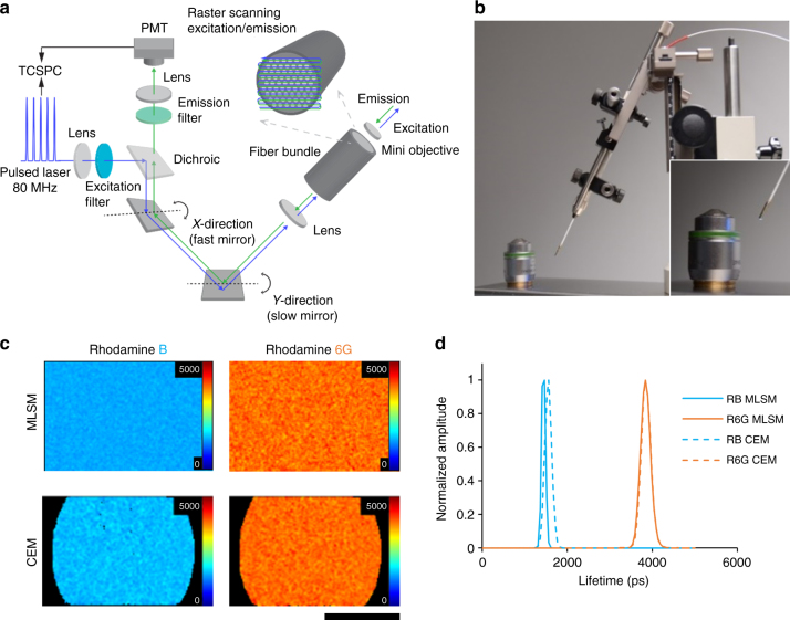
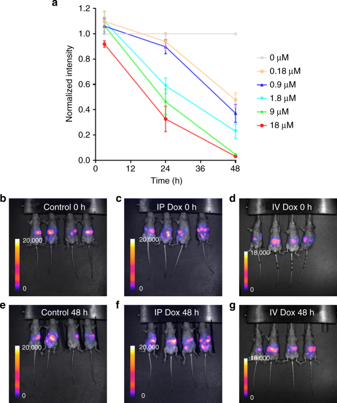
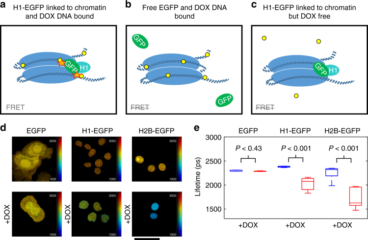
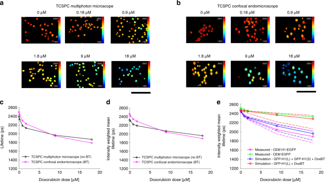
We present an approach to quantify drug-target engagement using in vivo fluorescence endomicroscopy, validated with in vitro measurements. Doxorubicin binding to chromatin changes the fluorescence lifetime of histone-GFP fusions that we measure in vivo at single-cell resolution using a confocal laparo/endomicroscope. We measure both intra- and inter-tumor heterogeneity in doxorubicin chromatin engagement in a model of peritoneal metastasis of ovarian cancer, revealing striking variation in the efficacy of doxorubicin-chromatin binding depending on intra-peritoneal or intravenous delivery. Further, we observe significant variations in doxorubicin-chromatin binding between different metastases in the same mouse and between different regions of the same metastasis. The quantitative nature of fluorescence lifetime imaging enables direct comparison of drug-target engagement for different drug delivery routes and between in vitro and in vivo experiments. This uncovers different rates of cell killing for the same level of doxorubicin binding in vitro and in vivo.
我们提出了一种使用体内荧光内窥技术定量药物-靶标结合的方法,并通过体外测量进行了验证。阿霉素与染色质结合会改变组蛋白-GFP 融合物的荧光寿命,我们使用共聚焦腹腔镜/内窥显微镜以单细胞分辨率在体内进行测量。我们在卵巢癌腹膜转移模型中测量了阿霉素与染色质结合的肿瘤内和肿瘤间异质性,结果显示,阿霉素与染色质结合的效果取决于腹腔内或静脉内给药方式,差异显著。此外,我们还观察到同一只小鼠的不同转移灶以及同一转移灶的不同区域之间,阿霉素与染色质的结合存在显著差异。荧光寿命成像的定量性质可以实现不同药物给药途径之间以及体外和体内实验之间的药物-靶标结合的直接比较。这揭示了相同水平的阿霉素结合在体外和体内的细胞杀伤率不同。