Department of Chemistry, The University of Tokyo, Tokyo, Japan.
Department of Neurosurgical Engineering and Translational Neuroscience, Tohoku University Graduate School of Medicine, Miyagi, Japan.
Nat Commun. 2024 Sep 4;15(1):7376. doi: 10.1038/s41467-024-51125-y.
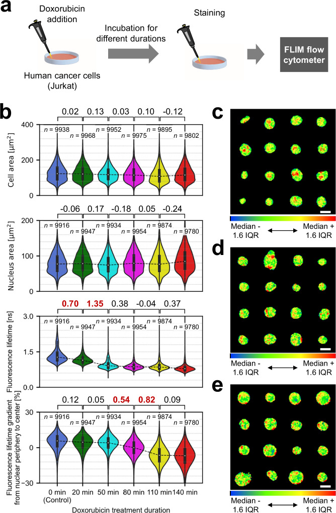
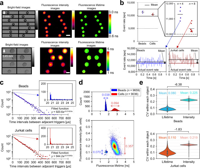
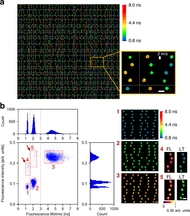
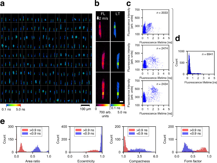
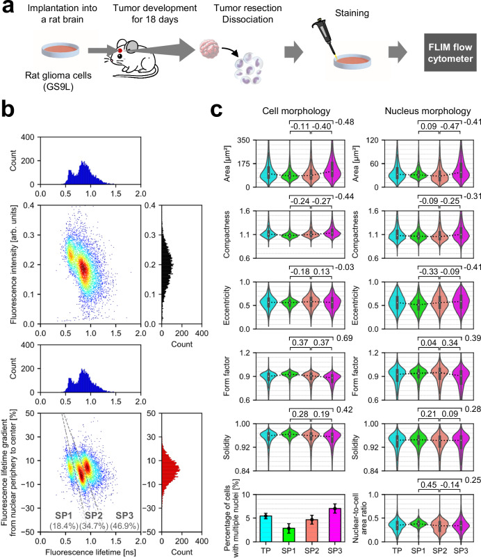
Flow cytometry is a vital tool in biomedical research and laboratory medicine. However, its accuracy is often compromised by undesired fluctuations in fluorescence intensity. While fluorescence lifetime imaging microscopy (FLIM) bypasses this challenge as fluorescence lifetime remains unaffected by such fluctuations, the full integration of FLIM into flow cytometry has yet to be demonstrated due to speed limitations. Here we overcome the speed limitations in FLIM, thereby enabling high-throughput FLIM flow cytometry at a high rate of over 10,000 cells per second. This is made possible by using dual intensity-modulated continuous-wave beam arrays with complementary modulation frequency pairs for fluorophore excitation and acquiring fluorescence lifetime images of rapidly flowing cells. Moreover, our FLIM system distinguishes subpopulations in male rat glioma and captures dynamic changes in the cell nucleus induced by an anti-cancer drug. FLIM flow cytometry significantly enhances cellular analysis capabilities, providing detailed insights into cellular functions, interactions, and environments.
流式细胞术是生物医学研究和实验室医学的重要工具。然而,其准确性经常受到荧光强度不期望波动的影响。尽管荧光寿命成像显微镜(FLIM)避免了这一挑战,因为荧光寿命不受这种波动的影响,但由于速度限制,FLIM 尚未完全集成到流式细胞术中。在这里,我们克服了 FLIM 的速度限制,从而能够以每秒超过 10000 个细胞的高通量进行 FLIM 流式细胞术。这是通过使用具有互补调制频率对的双强度调制连续波光束阵列来实现的,用于荧光团激发,并获取快速流动细胞的荧光寿命图像。此外,我们的 FLIM 系统能够区分雄性大鼠神经胶质瘤中的亚群,并捕获抗癌药物诱导的细胞核内的动态变化。FLIM 流式细胞术显著增强了细胞分析能力,提供了对细胞功能、相互作用和环境的详细了解。