Institute of Biochemistry and Microbiology, Faculty of Chemical and Food Technology, Slovak University of Technology in Bratislava, Radlinského 9, 812 37 Bratislava 1, Slovakia.
Institute of Molecular Physiology and Genetics, Centre of Bioscience, Slovak Academy of Sciences, Dúbravská cesta 9, 845 05 Bratislava 4, Slovakia.
Int J Mol Sci. 2018 Jul 7;19(7):1985. doi: 10.3390/ijms19071985.
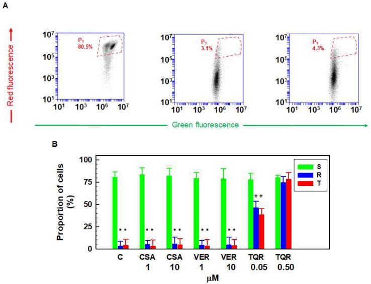
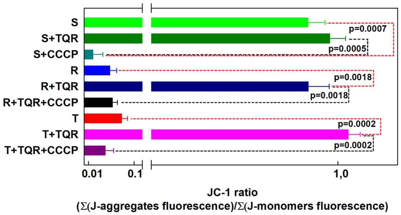
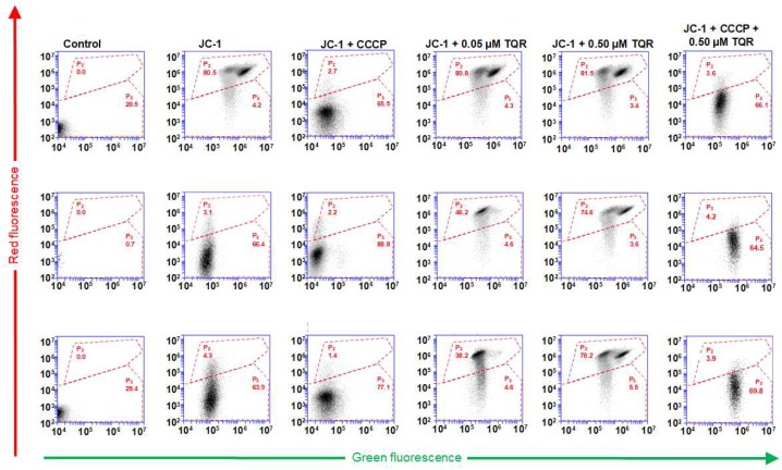
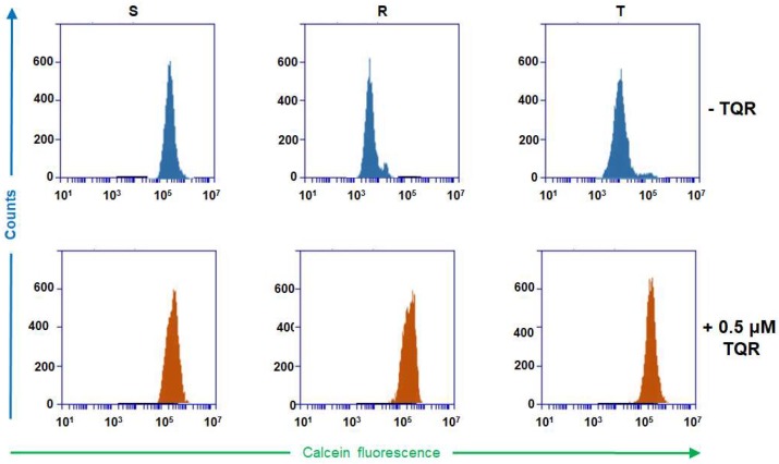
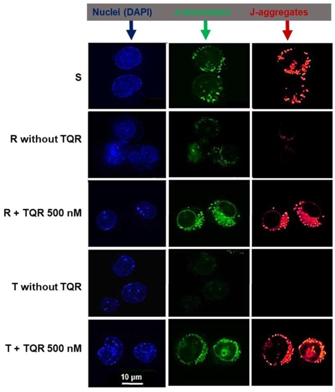
JC-1, a cationic fluorescent dye when added to living cells, is known to be localized exclusively in mitochondria, particularly in good physiological conditions characterized by sufficient mitochondrial membrane potential (ΔΨ). The accumulation of JC-1 in these organelles leads to the formation J-aggregates (with a specific red fluorescence emission maximum at 590 nm), which is in addition to the typical green fluorescence of J-monomers (emission maximum of ∼529 nm). The lack of mitochondrial ΔΨ leads to the depression of JC-1 mitochondrial accumulation and a decrease in J-aggregate formation. Therefore, the ratio between the red and green fluorescence of cells loaded with JC-1 is often used for the detection of the mitochondrial membrane potential. However, JC-1 represents a suitable substrate of the multidrug transporter P-glycoprotein (P-gp). Therefore, the depression of the JC-1 content in intracellular space and particularly in the mitochondria to a level that is inefficient for J-aggregate formation could be expected in P-gp-positive cells. In the current paper, we proved this behavior on parental P-gp-negative L1210 (S) cells and their P-gp-positive variants obtained by either selection with vincristine (R) or transfection with the human gene encoding P-gp (T). P-glycoprotein inhibitors cyclosporine A and verapamil fail to restore JC-1 loading of the R and T cells to an extent similar to that observed in S cells. In contrast, the noncompetitive high affinity P-gp inhibitor tariquidar fully restored JC-1 accumulation and the presence of the typical red fluorescence of J-aggregates. In the presence of tariquidar, measurement of the JC-1 fluorescence revealed similar levels of mitochondrial membrane potential in P-gp-negative (S) and P-gp-positive cells (R and T).
JC-1 是一种阳离子荧光染料,当添加到活细胞中时,已知其专门定位于线粒体中,尤其是在具有足够线粒体膜电位(ΔΨ)的良好生理条件下。JC-1 在这些细胞器中的积累导致 J-聚集体的形成(在 590nm 处具有特定的红色荧光发射最大值),这是 J-单体(约 529nm 的典型绿色荧光发射最大值)的补充。缺乏线粒体 ΔΨ 会导致 JC-1 在线粒体中的积累减少,J-聚集体的形成减少。因此,用 JC-1 加载的细胞的红色和绿色荧光的比率通常用于检测线粒体膜电位。然而,JC-1 是多药转运蛋白 P-糖蛋白(P-gp)的合适底物。因此,预计 P-gp 阳性细胞的细胞内空间,特别是在线粒体中,JC-1 的含量会降低到形成 J-聚集体效率低下的水平。在本文中,我们在亲本 P-gp 阴性 L1210(S)细胞及其通过长春新碱(R)选择或转染编码 P-gp 的人基因(T)获得的 P-gp 阳性变体上证明了这种行为。环孢素 A 和维拉帕米等 P-糖蛋白抑制剂未能使 R 和 T 细胞的 JC-1 负载恢复到与 S 细胞观察到的相似程度。相比之下,非竞争性高亲和力 P-gp 抑制剂他利喹达完全恢复了 JC-1 的积累以及 J-聚集体的典型红色荧光的存在。在他利喹达存在的情况下,JC-1 荧光的测量显示 P-gp 阴性(S)和 P-gp 阳性细胞(R 和 T)的线粒体膜电位相似。