Klotz Daniela, Hirzmann Jörg, Bauer Christian, Schöne Joachim, Iseringhausen Maximilian, Wohlsein Peter, Baumgärtner Wolfgang, Herder Vanessa
Department of Pathology, University of Veterinary Medicine Hannover, Hannover, Germany.
Institute of Parasitology, Justus Liebig University, Giessen, Germany.
Int J Parasitol Parasites Wildl. 2018 Mar 2;7(1):99-105. doi: 10.1016/j.ijppaw.2018.02.003. eCollection 2018 Apr.
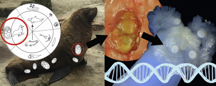
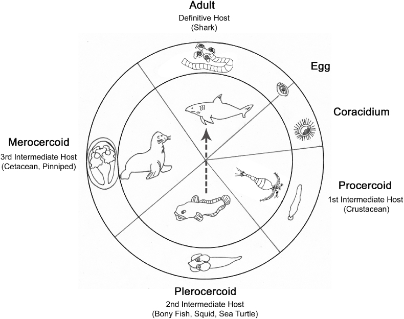
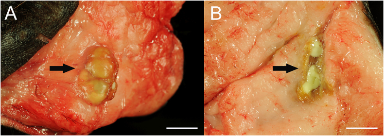
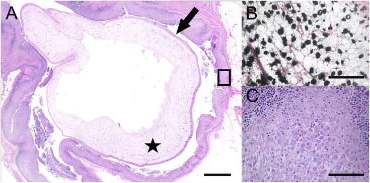
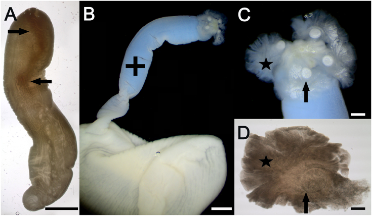
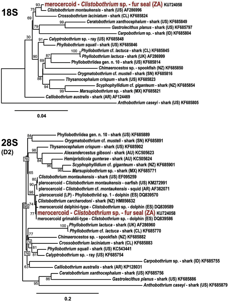
Fur seals represent intermediate hosts of the cestode . Large sharks are definitive hosts for these parasites. Two female, 25- and 27-year-old fur seals, caught in the 1980s at the South African coast, were examined pathomorphologically. Both animals showed multifocal, up to 1 cm in diameter large cavities of the thoracic and abdominal subcutaneous adipose tissue containing intraluminal metacestodes of tapeworms, which were surrounded by a locally extensive, pyogranulomatous panniculitis. The metacestodes (merocercoids) of one fur seal were isolated from the subcutaneous adipose tissue and characterized morphologically and for the first time from this host by molecular techniques. The morphometric data corresponded with 'delphini'-morphotype merocercoids, but the sequence of the partial 28S ribosomal RNA gene identified them as conspecific with merocercoids of the morphotype 'grimaldii. These merocercoid types are morphologically Type XV metacestodes of marine tapeworms and represent different species of . Sequence data were generated for 18S, ITS1, 5.8S, ITS2, partial 28S ribosomal DNA and partial mitochondrial cox1 gene and phylogenetic analysis of 18S rRNA and partial 28S rRNA genes identified the fur seal merocercoids as species. However, it cannot yet be assigned to species level because of limited molecular data from adult stages. Most likely, both fur seals were infected as juveniles in their original habitat, the coastal regions of South Africa. The metacestode infection is probably an incidental finding, however, there is a chronic inflammatory reaction next to the subcutaneous merocercoids. It is noteworthy, that the merocercoids remain in a potentially infective stage even after more than 20 years.
海狗是该绦虫的中间宿主。大型鲨鱼是这些寄生虫的终宿主。对20世纪80年代在南非海岸捕获的两只分别为25岁和27岁的雌性海狗进行了病理形态学检查。两只动物的胸腹部皮下脂肪组织均出现多灶性、直径达1厘米的大腔隙,腔内含有绦虫的囊尾蚴,周围有局部广泛的脓性肉芽肿性脂膜炎。从一只海狗的皮下脂肪组织中分离出囊尾蚴(豆状囊尾蚴),并首次通过分子技术对其进行形态学鉴定。形态测量数据与“delphini”形态型豆状囊尾蚴相符,但部分28S核糖体RNA基因序列将它们鉴定为与“grimaldii”形态型豆状囊尾蚴同种。这些豆状囊尾蚴类型在形态上是海洋绦虫的XV型囊尾蚴,代表不同的物种。生成了18S、ITS1、5.8S、ITS2、部分28S核糖体DNA和部分线粒体cox1基因的序列数据,对18S rRNA和部分28S rRNA基因的系统发育分析将海狗豆状囊尾蚴鉴定为 物种。然而,由于来自成虫阶段的分子数据有限,目前尚不能将其归为物种水平。很可能这两只海狗在其原生栖息地南非沿海地区幼年时就受到了感染。囊尾蚴感染可能是一个偶然发现,不过,皮下豆状囊尾蚴旁有慢性炎症反应。值得注意的是,即使经过20多年,豆状囊尾蚴仍处于潜在感染阶段。