• 文献检索
  • 文档翻译
  • 深度研究
  • 学术资讯
  • Suppr Zotero 插件Zotero 插件
  • 邀请有礼
  • 套餐&价格
  • 历史记录
应用&插件
Suppr Zotero 插件Zotero 插件浏览器插件Mac 客户端Windows 客户端微信小程序
定价
高级版会员购买积分包购买API积分包
服务
文献检索文档翻译深度研究API 文档MCP 服务
关于我们
关于 Suppr公司介绍联系我们用户协议隐私条款
关注我们

Suppr 超能文献

核心技术专利:CN118964589B侵权必究
粤ICP备2023148730 号-1Suppr @ 2026

文献检索

告别复杂PubMed语法,用中文像聊天一样搜索,搜遍4000万医学文献。AI智能推荐,让科研检索更轻松。

立即免费搜索

文件翻译

保留排版,准确专业,支持PDF/Word/PPT等文件格式,支持 12+语言互译。

免费翻译文档

深度研究

AI帮你快速写综述,25分钟生成高质量综述,智能提取关键信息,辅助科研写作。

立即免费体验

相似文献

1
Microbubble Localization for Three-Dimensional Superresolution Ultrasound Imaging Using Curve Fitting and Deconvolution Methods.基于曲线拟合和反卷积方法的微泡三维超分辨超声成像的微泡定位。
IEEE Trans Biomed Eng. 2018 Dec;65(12):2692-2703. doi: 10.1109/TBME.2018.2813759. Epub 2018 Mar 8.
2
A dual-mode hemispherical sparse array for 3D passive acoustic mapping and skull localization within a clinical MRI guided focused ultrasound device.一种双模半球形稀疏阵,用于临床 MRI 引导聚焦超声设备内的 3D 被动声纳测绘和颅骨定位。
Phys Med Biol. 2018 Mar 15;63(6):065008. doi: 10.1088/1361-6560/aab0aa.
3
Experimental demonstration of passive acoustic imaging in the human skull cavity using CT-based aberration corrections.基于CT的像差校正技术在人类颅腔内进行被动声学成像的实验演示。
Med Phys. 2015 Jul;42(7):4385-400. doi: 10.1118/1.4922677.
4
Three-dimensional super resolution ultrasound imaging with a multi-frequency hemispherical phased array.三维超分辨率超声成像的多频半球形相控阵。
Med Phys. 2023 Dec;50(12):7478-7497. doi: 10.1002/mp.16733. Epub 2023 Sep 13.
5
A multi-frequency sparse hemispherical ultrasound phased array for microbubble-mediated transcranial therapy and simultaneous cavitation mapping.一种用于微泡介导的经颅治疗和同步空化映射的多频稀疏半球形超声相控阵。
Phys Med Biol. 2016 Dec 21;61(24):8476-8501. doi: 10.1088/0031-9155/61/24/8476. Epub 2016 Nov 15.
6
Ultrafast 3D Ultrasound Localization Microscopy Using a 32 × 32 Matrix Array.使用 32×32 矩阵阵元的超快 3D 超声定位显微镜。
IEEE Trans Med Imaging. 2019 Sep;38(9):2005-2015. doi: 10.1109/TMI.2018.2890358. Epub 2019 Apr 1.
7
Three-dimensional transcranial ultrasound imaging of microbubble clouds using a sparse hemispherical array.使用稀疏半球形阵列对微泡云进行三维经颅超声成像。
IEEE Trans Biomed Eng. 2014 Apr;61(4):1285-94. doi: 10.1109/TBME.2014.2300838.
8
3-D In Vitro Acoustic Super-Resolution and Super-Resolved Velocity Mapping Using Microbubbles.基于微泡的三维体外声超分辨率与超分辨率速度成像。
IEEE Trans Ultrason Ferroelectr Freq Control. 2017 Oct;64(10):1478-1486. doi: 10.1109/TUFFC.2017.2731664. Epub 2017 Jul 31.
9
Doppler Passive Acoustic Mapping.多普勒被动声纳测绘。
IEEE Trans Ultrason Ferroelectr Freq Control. 2020 Dec;67(12):2692-2703. doi: 10.1109/TUFFC.2020.3011657. Epub 2020 Nov 24.
10
On the Effects of Spatial Sampling Quantization in Super-Resolution Ultrasound Microvessel Imaging.超分辨率超声微血管成像中空间采样量化的影响。
IEEE Trans Ultrason Ferroelectr Freq Control. 2018 Dec;65(12):2264-2276. doi: 10.1109/TUFFC.2018.2832600. Epub 2018 May 4.

引用本文的文献

1
Advancements in Three-Dimensional Super-Resolution Ultrasound Imaging: A Narrative Review.三维超分辨率超声成像的进展:一项叙述性综述。
J Ultrasound Med. 2025 Jul;44(7):1157-1174. doi: 10.1002/jum.16682. Epub 2025 Mar 12.
2
Improving sub-pixel accuracy in ultrasound localization microscopy using supervised and self-supervised deep learning.使用监督式和自监督式深度学习提高超声定位显微镜中的亚像素精度。
Meas Sci Technol. 2024 Apr 1;35(4):045701. doi: 10.1088/1361-6501/ad1671. Epub 2024 Jan 9.
3
Three-dimensional super resolution ultrasound imaging with a multi-frequency hemispherical phased array.三维超分辨率超声成像的多频半球形相控阵。
Med Phys. 2023 Dec;50(12):7478-7497. doi: 10.1002/mp.16733. Epub 2023 Sep 13.
4
Real-Time Passive Acoustic Mapping With Enhanced Spatial Resolution in Neuronavigation-Guided Focused Ultrasound for Blood-Brain Barrier Opening.神经导航引导聚焦超声实时被动声纳成像提高血脑屏障开放的空间分辨率
IEEE Trans Biomed Eng. 2023 Oct;70(10):2874-2885. doi: 10.1109/TBME.2023.3266952. Epub 2023 Sep 27.
5
Detecting Early Ocular Choroidal Melanoma Using Ultrasound Localization Microscopy.使用超声定位显微镜检测早期眼部脉络膜黑色素瘤。
Bioengineering (Basel). 2023 Mar 28;10(4):428. doi: 10.3390/bioengineering10040428.
6
Fast 3D Super-Resolution Ultrasound With Adaptive Weight-Based Beamforming.基于自适应权重的波束形成的快速三维超分辨率超声。
IEEE Trans Biomed Eng. 2023 Sep;70(9):2752-2761. doi: 10.1109/TBME.2023.3263369. Epub 2023 Aug 30.
7
Cerebral microcirculation mapped by echo particle tracking velocimetry quantifies the intracranial pressure and detects ischemia.应用示踪微泡速度描记技术对脑微循环进行映射,可定量测量颅内压并检测缺血情况。
Nat Commun. 2022 Feb 3;13(1):666. doi: 10.1038/s41467-022-28298-5.
8
In Vivo Visualization of Eye Vasculature Using Super-Resolution Ultrasound Microvessel Imaging.利用超分辨率超声微血管成像技术对眼部血管进行体内可视化。
IEEE Trans Biomed Eng. 2020 Oct;67(10):2870-2880. doi: 10.1109/TBME.2020.2972514. Epub 2020 Feb 10.
9
Super-resolution Ultrasound Imaging.超分辨率超声成像。
Ultrasound Med Biol. 2020 Apr;46(4):865-891. doi: 10.1016/j.ultrasmedbio.2019.11.013. Epub 2020 Jan 21.
10
Computed Tomography (CT) Image Quality Enhancement via a Uniform Framework Integrating Noise Estimation and Super-Resolution Networks.通过集成噪声估计和超分辨率网络的统一框架增强计算机断层扫描(CT)图像质量
Sensors (Basel). 2019 Jul 30;19(15):3348. doi: 10.3390/s19153348.

本文引用的文献

1
Ultrasound localization microscopy to image and assess microvasculature in a rat kidney.超声定位显微镜成像和评估大鼠肾脏的微血管。
Sci Rep. 2017 Oct 20;7(1):13662. doi: 10.1038/s41598-017-13676-7.
2
Subwavelength motion-correction for ultrafast ultrasound localization microscopy.用于超快超声定位显微镜的亚波长运动校正
Ultrasonics. 2017 May;77:17-21. doi: 10.1016/j.ultras.2017.01.008. Epub 2017 Jan 18.
3
A multi-frequency sparse hemispherical ultrasound phased array for microbubble-mediated transcranial therapy and simultaneous cavitation mapping.一种用于微泡介导的经颅治疗和同步空化映射的多频稀疏半球形超声相控阵。
Phys Med Biol. 2016 Dec 21;61(24):8476-8501. doi: 10.1088/0031-9155/61/24/8476. Epub 2016 Nov 15.
4
Ultrafast ultrasound localization microscopy for deep super-resolution vascular imaging.超快超声定位显微镜用于深层超分辨率血管成像。
Nature. 2015 Nov 26;527(7579):499-502. doi: 10.1038/nature16066.
5
Detection and Tracking of Multiple Microbubbles in Ultrasound B-Mode Images.超声B模式图像中多个微泡的检测与跟踪
IEEE Trans Ultrason Ferroelectr Freq Control. 2016 Jan;63(1):72-82. doi: 10.1109/TUFFC.2015.2500266. Epub 2015 Nov 17.
6
Resolution limits of ultrafast ultrasound localization microscopy.超快超声定位显微镜的分辨率极限
Phys Med Biol. 2015 Nov 21;60(22):8723-40. doi: 10.1088/0031-9155/60/22/8723. Epub 2015 Oct 28.
7
A high-density 3D localization algorithm for stochastic optical reconstruction microscopy.一种用于随机光学重建显微镜的高密度三维定位算法。
Opt Nanoscopy. 2012;1(6). doi: 10.1186/2192-2853-1-6.
8
In vivo acoustic super-resolution and super-resolved velocity mapping using microbubbles.利用微泡进行体内声超分辨率和超分辨率速度成像。
IEEE Trans Med Imaging. 2015 Feb;34(2):433-40. doi: 10.1109/TMI.2014.2359650. Epub 2014 Sep 23.
9
Three-dimensional transcranial ultrasound imaging of microbubble clouds using a sparse hemispherical array.使用稀疏半球形阵列对微泡云进行三维经颅超声成像。
IEEE Trans Biomed Eng. 2014 Apr;61(4):1285-94. doi: 10.1109/TBME.2014.2300838.
10
A super-resolution ultrasound method for brain vascular mapping.一种用于脑血管测绘的超高分辨率超声方法。
Med Phys. 2013 Nov;40(11):110701. doi: 10.1118/1.4823762.

基于曲线拟合和反卷积方法的微泡三维超分辨超声成像的微泡定位。

Microbubble Localization for Three-Dimensional Superresolution Ultrasound Imaging Using Curve Fitting and Deconvolution Methods.

出版信息

IEEE Trans Biomed Eng. 2018 Dec;65(12):2692-2703. doi: 10.1109/TBME.2018.2813759. Epub 2018 Mar 8.

DOI:10.1109/TBME.2018.2813759
PMID:29993387
原文链接:https://pmc.ncbi.nlm.nih.gov/articles/PMC6459186/
Abstract

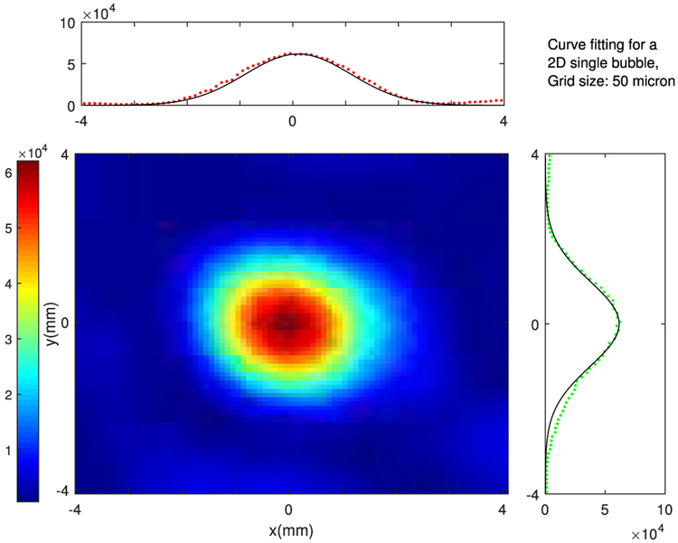
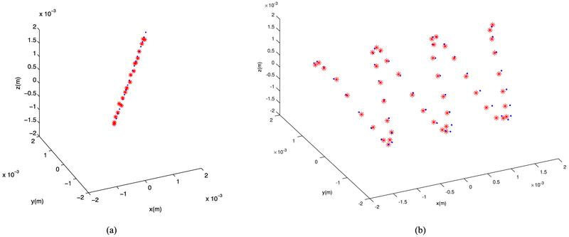
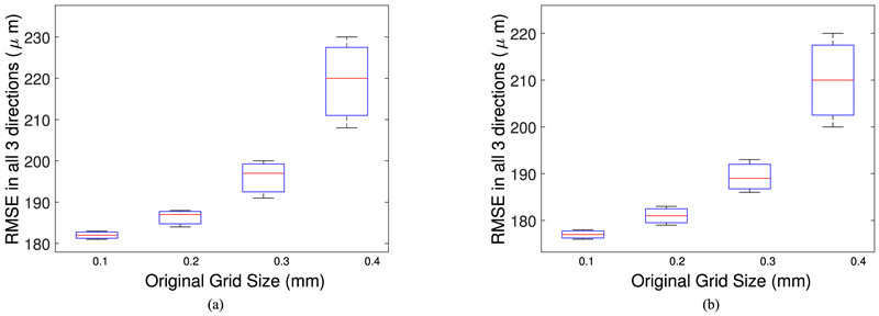
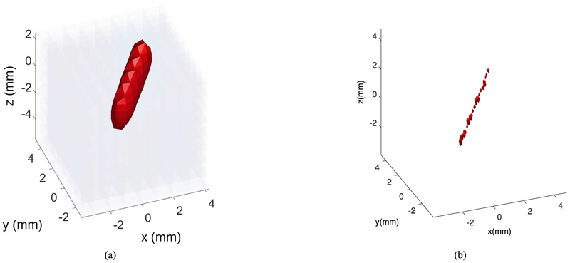
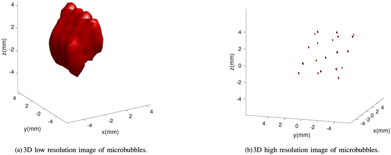
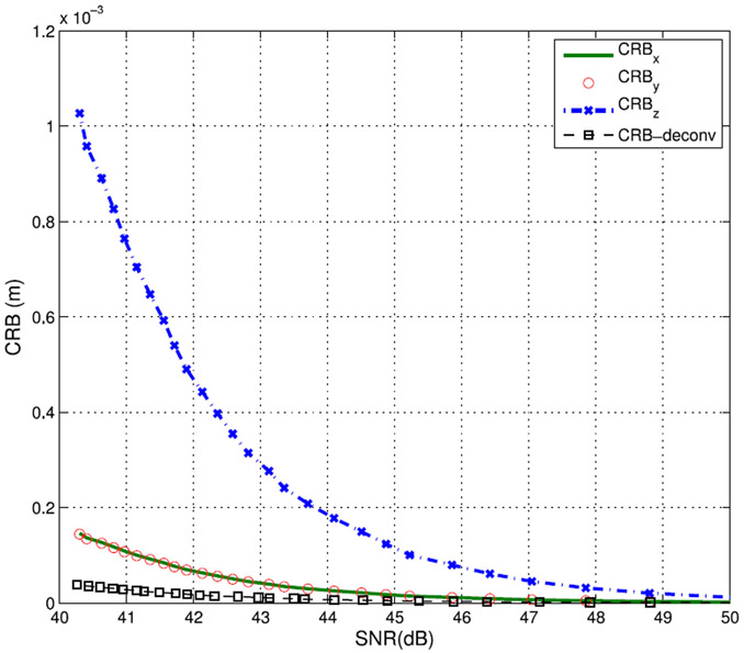
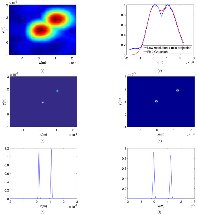
Superresolution algorithms in ultrasound imaging are attracting the interest of researchers recently due to the ability of these methods to enable enhanced vascular imaging. In this study, two superresolution imaging methods are compared for postprocessing images of microbubbles generated using passive acoustic mapping (PAM) methods with a potential application of three-dimensional (3-D) brain vascular imaging. The first method is based on fitting single bubble images one at a time with a 3-D Gaussian profile to localize the microbubbles and a superresolution image is then formed using the uncertainty of the localization as the standard deviation of the Gaussian profile. The second superresolution method is based on image deconvolution that processes multiframe resolution-limited images iteratively and estimates the intensity at each pixel of the superresolution image without the need for localizing each microbubble. The point spread function is approximated by a Gaussian curve which is similar to the beam response of the hemispherical transducer array used in our experimental setup. The Cramér-Rao Bounds of the two estimation techniques are derived analytically and the performance of these techniques is compared through numerical simulations based on experimental PAM images. For linear and sinusoidal traces, the localization errors between the estimated peaks by the fitting-based method and the actual source locations were 220 10 m and 210 5 m, respectively, as compared to 74 10 m and 59 8 m with the deconvolution-based method. However, in terms of the running time and the computational costs, the curve fitting technique outperforms the deconvolution-based approach.

摘要

超声成像中的超分辨率算法最近引起了研究人员的兴趣,因为这些方法能够增强血管成像。在这项研究中,比较了两种超分辨率成像方法,用于处理使用被动声学映射 (PAM) 方法生成的微泡的后处理图像,潜在应用于三维 (3-D) 脑血管成像。第一种方法基于逐个拟合单个气泡图像的 3-D 高斯分布来定位微泡,然后使用定位的不确定性作为高斯分布的标准差来形成超分辨率图像。第二种超分辨率方法基于图像反卷积,它迭代地处理多帧分辨率受限的图像,并在不需要定位每个微泡的情况下估计超分辨率图像中每个像素的强度。点扩散函数通过高斯曲线近似,该曲线类似于我们实验设置中使用的半球形换能器阵列的波束响应。两种估计技术的克拉美罗界 (Cramér-Rao Bounds) 从理论上推导出来,并通过基于实验 PAM 图像的数值模拟比较了这些技术的性能。对于线性和正弦迹线,基于拟合的方法估计的峰值与实际源位置之间的定位误差分别为 220 10 m 和 210 5 m,而基于反卷积的方法分别为 74 10 m 和 59 8 m。然而,就运行时间和计算成本而言,曲线拟合技术优于基于反卷积的方法。

https://cdn.ncbi.nlm.nih.gov/pmc/blobs/26aa/6459186/cb805390d4c6/nihms-1020880-f0011.jpg
https://cdn.ncbi.nlm.nih.gov/pmc/blobs/26aa/6459186/56b7ab66179e/nihms-1020880-f0001.jpg
https://cdn.ncbi.nlm.nih.gov/pmc/blobs/26aa/6459186/e5c43b0bf945/nihms-1020880-f0002.jpg
https://cdn.ncbi.nlm.nih.gov/pmc/blobs/26aa/6459186/0837d3f0c66f/nihms-1020880-f0003.jpg
https://cdn.ncbi.nlm.nih.gov/pmc/blobs/26aa/6459186/37ef98565bb2/nihms-1020880-f0004.jpg
https://cdn.ncbi.nlm.nih.gov/pmc/blobs/26aa/6459186/114be4495468/nihms-1020880-f0005.jpg
https://cdn.ncbi.nlm.nih.gov/pmc/blobs/26aa/6459186/9561419fbadb/nihms-1020880-f0006.jpg
https://cdn.ncbi.nlm.nih.gov/pmc/blobs/26aa/6459186/e0d642246145/nihms-1020880-f0007.jpg
https://cdn.ncbi.nlm.nih.gov/pmc/blobs/26aa/6459186/aef4b6da642c/nihms-1020880-f0008.jpg
https://cdn.ncbi.nlm.nih.gov/pmc/blobs/26aa/6459186/0a41d39d519a/nihms-1020880-f0009.jpg
https://cdn.ncbi.nlm.nih.gov/pmc/blobs/26aa/6459186/4209603f4cf0/nihms-1020880-f0010.jpg
https://cdn.ncbi.nlm.nih.gov/pmc/blobs/26aa/6459186/cb805390d4c6/nihms-1020880-f0011.jpg