1 Plastic and Reconstructive Surgery, Yale University School of Medicine, Yale University, New Haven, CT, USA.
2 Department of Materials Science and Engineering, McCormick School of Engineering, Northwestern University, Chicago, IL, USA.
Cell Transplant. 2018 Aug;27(8):1269-1280. doi: 10.1177/0963689718782452. Epub 2018 Jul 16.
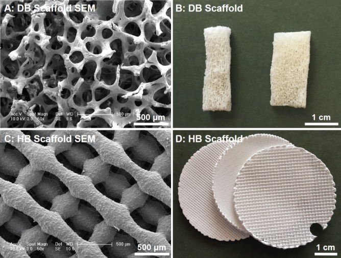
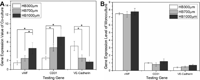
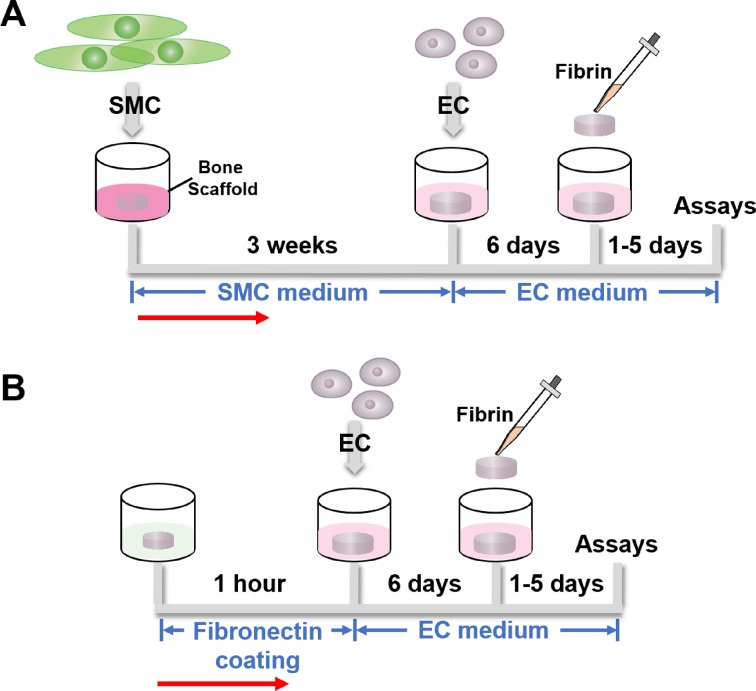
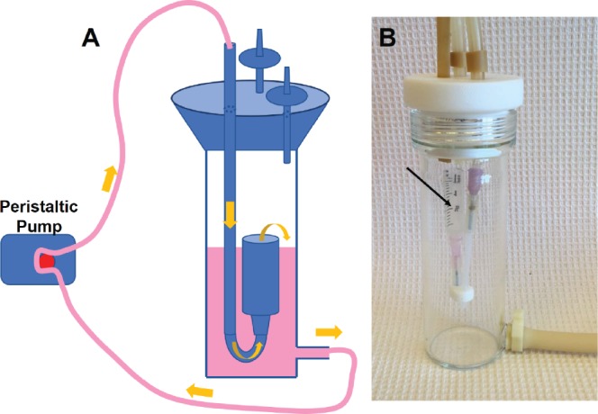
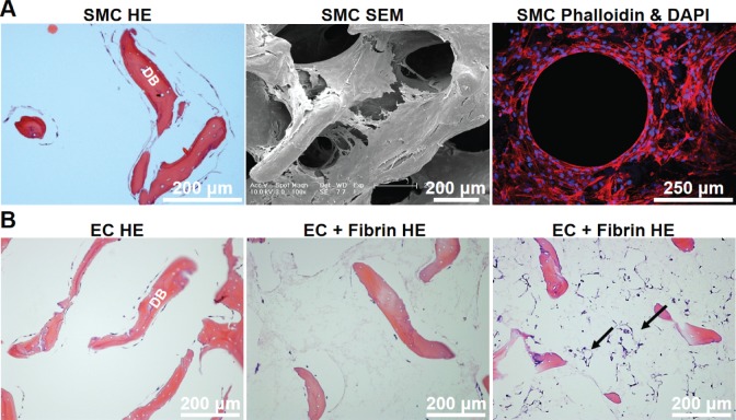
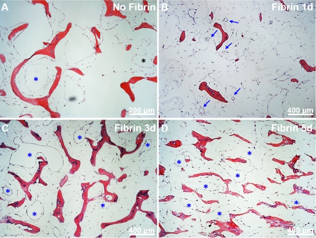
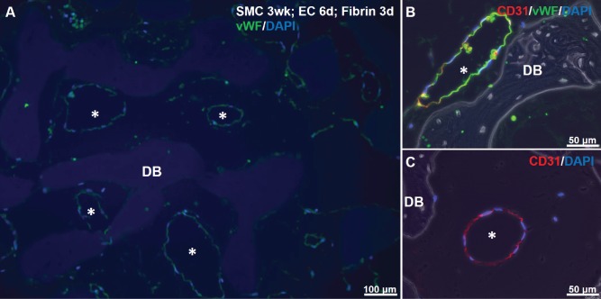
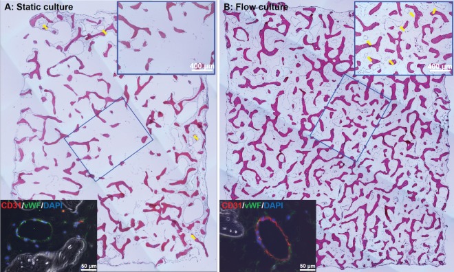
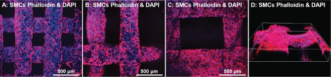
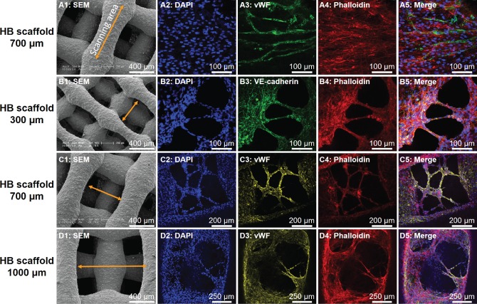
Vascularization of engineered bone tissue is critical for ensuring its survival after implantation. In vitro pre-vascularization of bone grafts with endothelial cells is a promising strategy to improve implant survival. In this study, we pre-cultured human smooth muscle cells (hSMCs) on bone scaffolds for 3 weeks followed by seeding of human umbilical vein endothelial cells (HUVECs), which produced a desirable environment for microvasculature formation. The sequential cell-seeding protocol was successfully applied to both natural (decellularized native bone, or DB) and synthetic (3D-printed Hyperelastic "Bone" scaffolds, or HB) scaffolds, demonstrating a comprehensive platform for developing natural and synthetic-based in vitro vascularized bone grafts. Using this sequential cell-seeding process, the HUVECs formed lumen structures throughout the DB scaffolds as well as vascular tissue bridging 3D-printed fibers within the HB. The pre-cultured hSMCs were essential for endothelial cell (EC) lumen formation within DB scaffolds, as well as for upregulating EC-specific gene expression of HUVECs grown on HB scaffolds. We further applied this co-culture protocol to DB scaffolds using a perfusion bioreactor, to overcome the limitations of diffusive mass transport into the interiors of the scaffolds. Compared with static culture, panoramic histological sections of DB scaffolds cultured in bioreactors showed improved cellular density, as well as a nominal increase in the number of lumen structures formed by ECs in the interior regions of the scaffolds. In conclusion, we have demonstrated that the sequential seeding of hSMCs and HUVECs can serve to generate early microvascular networks that could further support the in vitro tissue engineering of naturally or synthetically derived bone grafts and in both random (DB) and ordered (HB) pore networks. Combined with the preliminary bioreactor study, this process also shows potential to generate clinically sized, vascularized bone scaffolds for tissue and regenerative engineering.
工程化骨组织的血管化对于确保其植入后的存活至关重要。将内皮细胞预先血管化骨移植物是提高植入物存活率的一种有前途的策略。在这项研究中,我们将人平滑肌细胞(hSMC)预培养在骨支架上 3 周,然后接种人脐静脉内皮细胞(HUVEC),从而为微血管形成创造了理想的环境。该顺序细胞接种方案成功应用于天然(去细胞化天然骨,或 DB)和合成(3D 打印超弹性“骨”支架,或 HB)支架,为开发天然和合成的体外血管化骨移植物提供了一个全面的平台。使用这种顺序细胞接种过程,HUVEC 在 DB 支架内形成了腔结构,并在 HB 内的 3D 打印纤维内形成了血管组织桥接。预培养的 hSMC 对于 DB 支架内内皮细胞(EC)腔结构的形成以及对在 HB 支架上生长的 HUVEC 的 EC 特异性基因表达的上调至关重要。我们进一步将这种共培养方案应用于使用灌注生物反应器的 DB 支架,以克服扩散质量传输到支架内部的限制。与静态培养相比,在生物反应器中培养的 DB 支架的全景组织学切片显示出改善的细胞密度,以及 EC 在支架内部区域形成的腔结构数量的名义增加。总之,我们已经证明,hSMC 和 HUVEC 的顺序接种可以产生早期的微血管网络,这可以进一步支持天然或合成衍生的骨移植物的体外组织工程,以及随机(DB)和有序(HB)孔网络。结合初步的生物反应器研究,该过程也显示出用于组织和再生工程的临床规模血管化骨支架的潜力。