Dos Santos Isabela B, da Silva Danielle A M, Paz Fabiana A C R, Garcia Daniel M, Carmona Adriana K, Teixeira Daniela, Longo-Maugéri Ieda M, Katz Simone, Barbiéri Clara L
Departamento de Microbiologia, Imunologia e Parasitologia, Escola Paulista de Medicina, Universidade Federal de São Paulo, São Paulo, Brazil.
Departamento de Farmacologia, Escola Paulista de Medicina, Universidade Federal de São Paulo, São Paulo, Brazil.
Front Microbiol. 2018 Jul 3;9:1427. doi: 10.3389/fmicb.2018.01427. eCollection 2018.
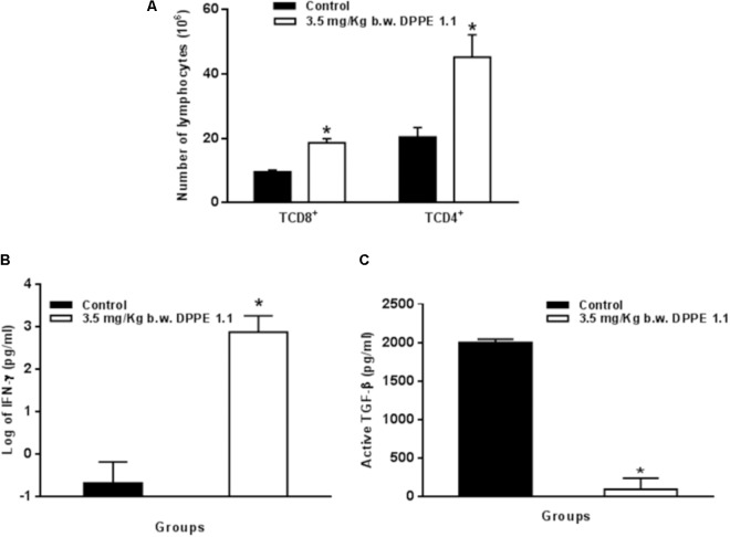
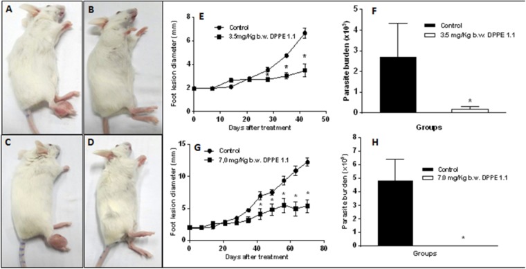
The present study focused on the activity of the palladacycle complex DPPE 1.1 on . Promastigotes of were destroyed by nanomolar concentrations of DPPE 1.1, whereas intracellular amastigotes were killed at drug concentrations fivefold less toxic than those harmful to macrophages. -infected BALB/c mice were treated by intralesional injection of DPPE 1.1. Animals treated with 3.5 and 7.0 mg/kg of DPPE 1.1 showed a significant decrease of foot lesion sizes and a parasite load reduction of 93 and 99%, respectively, when compared to untreated controls. Furthermore, DPPE 1.1 was non-toxic to treated animals. The cathepsin B activity of amastigotes was inhibited by DPPE 1.1 as demonstrated spectrofluorometrically by use of a specific fluorogenic substrate. Analysis of T-cells populations in mice treated with DPPE 1.1 and untreated controls was performed by fluorescence-activated cell sorter (FACS). IFN-γ was measured in supernatants of lymphocytes from popliteal and inguinal lymph nodes isolated from treated and untreated mice and stimulated with extract and active TGF-β was evaluated in supernatants of foot lesions; both dosages were carried out by means of a double-sandwich ELISA assay. A significant increase of TCD4 and TCD8 lymphocytes and IFN-γ secretion was displayed in mice treated with DPPE 1.1 compared to untreated animals, whereas a significant reduction of active TGF-β was observed in treated mice. These findings open perspectives for further investment in DPPE 1.1 as an alternative option for the chemotherapy of cutaneous leishmaniasis.
本研究聚焦于钯环配合物DPPE 1.1的活性。纳米摩尔浓度的DPPE 1.1可破坏 的前鞭毛体,而细胞内无鞭毛体在药物浓度下被杀死,该浓度对巨噬细胞的毒性比有害浓度低五倍。用DPPE 1.1病灶内注射治疗感染 的BALB/c小鼠。与未治疗的对照组相比,用3.5和7.0 mg/kg的DPPE 1.1治疗的动物足部病变大小显著减小,寄生虫负荷分别降低了93%和99%。此外,DPPE 1.1对治疗的动物无毒。通过使用特定的荧光底物进行荧光分光光度法证明,DPPE 1.1可抑制 无鞭毛体的组织蛋白酶B活性。用荧光激活细胞分选仪(FACS)对用DPPE 1.1治疗的小鼠和未治疗的对照组的T细胞群体进行分析。在用提取物刺激后,从治疗和未治疗的小鼠分离的腘窝和腹股沟淋巴结淋巴细胞上清液中测量IFN-γ,并在足部病变上清液中评估活性TGF-β;两种剂量均通过双夹心ELISA测定法进行。与未治疗的动物相比,用DPPE 1.1治疗的小鼠显示TCD4和TCD8淋巴细胞以及IFN-γ分泌显著增加,而在治疗的小鼠中观察到活性TGF-β显著降低。这些发现为进一步研究DPPE 1.1作为皮肤利什曼病化疗的替代选择开辟了前景。