• 文献检索
  • 文档翻译
  • 深度研究
  • 学术资讯
  • Suppr Zotero 插件Zotero 插件
  • 邀请有礼
  • 套餐&价格
  • 历史记录
应用&插件
Suppr Zotero 插件Zotero 插件浏览器插件Mac 客户端Windows 客户端微信小程序
定价
高级版会员购买积分包购买API积分包
服务
文献检索文档翻译深度研究API 文档MCP 服务
关于我们
关于 Suppr公司介绍联系我们用户协议隐私条款
关注我们

Suppr 超能文献

核心技术专利:CN118964589B侵权必究
粤ICP备2023148730 号-1Suppr @ 2026

文献检索

告别复杂PubMed语法,用中文像聊天一样搜索,搜遍4000万医学文献。AI智能推荐,让科研检索更轻松。

立即免费搜索

文件翻译

保留排版,准确专业,支持PDF/Word/PPT等文件格式,支持 12+语言互译。

免费翻译文档

深度研究

AI帮你快速写综述,25分钟生成高质量综述,智能提取关键信息,辅助科研写作。

立即免费体验

胶质瘤干细胞的分子亚型和分化程序作为细胞外囊泡谱和内皮细胞刺激活性的决定因素

Molecular subtypes and differentiation programmes of glioma stem cells as determinants of extracellular vesicle profiles and endothelial cell-stimulating activities.

作者信息

Spinelli C, Montermini L, Meehan B, Brisson A R, Tan S, Choi D, Nakano I, Rak J

机构信息

Department of Pediatrics, McGill University, The Research Institute of the McGill University Health Centre, Montreal, Canada.

UMR-CBMN CNRS, University of Bordeaux, IPB, France.

出版信息

J Extracell Vesicles. 2018 Jul 17;7(1):1490144. doi: 10.1080/20013078.2018.1490144. eCollection 2018.

DOI:10.1080/20013078.2018.1490144
PMID:30034643
原文链接:https://pmc.ncbi.nlm.nih.gov/articles/PMC6052423/
Abstract

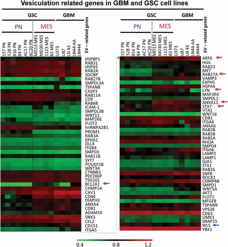
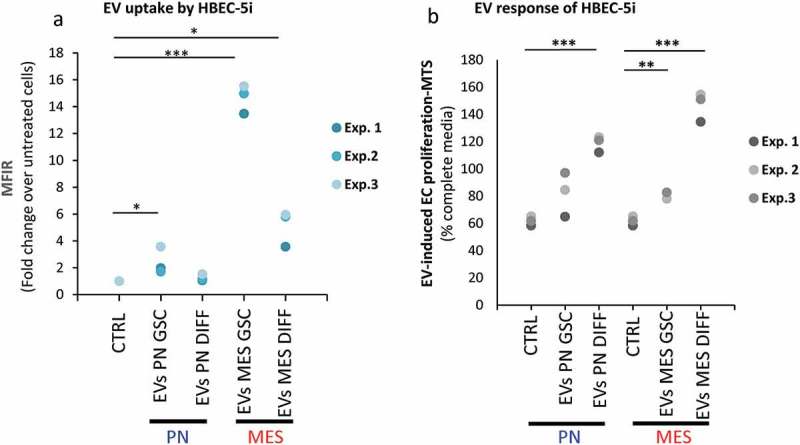
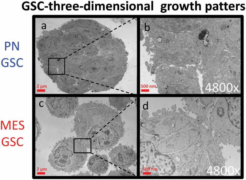
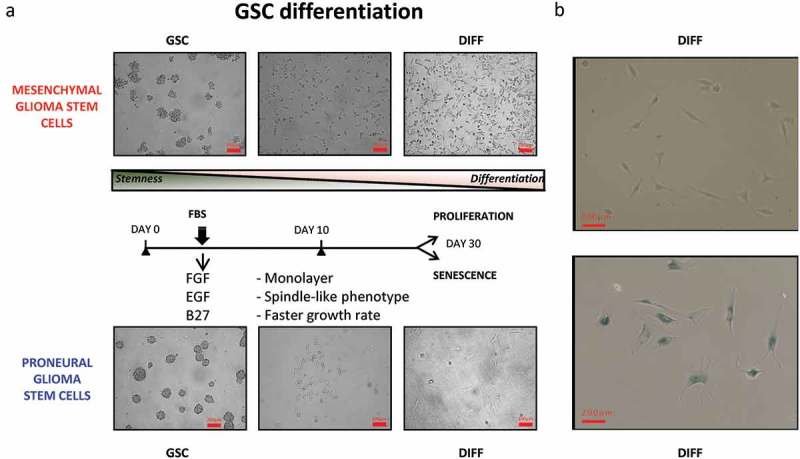
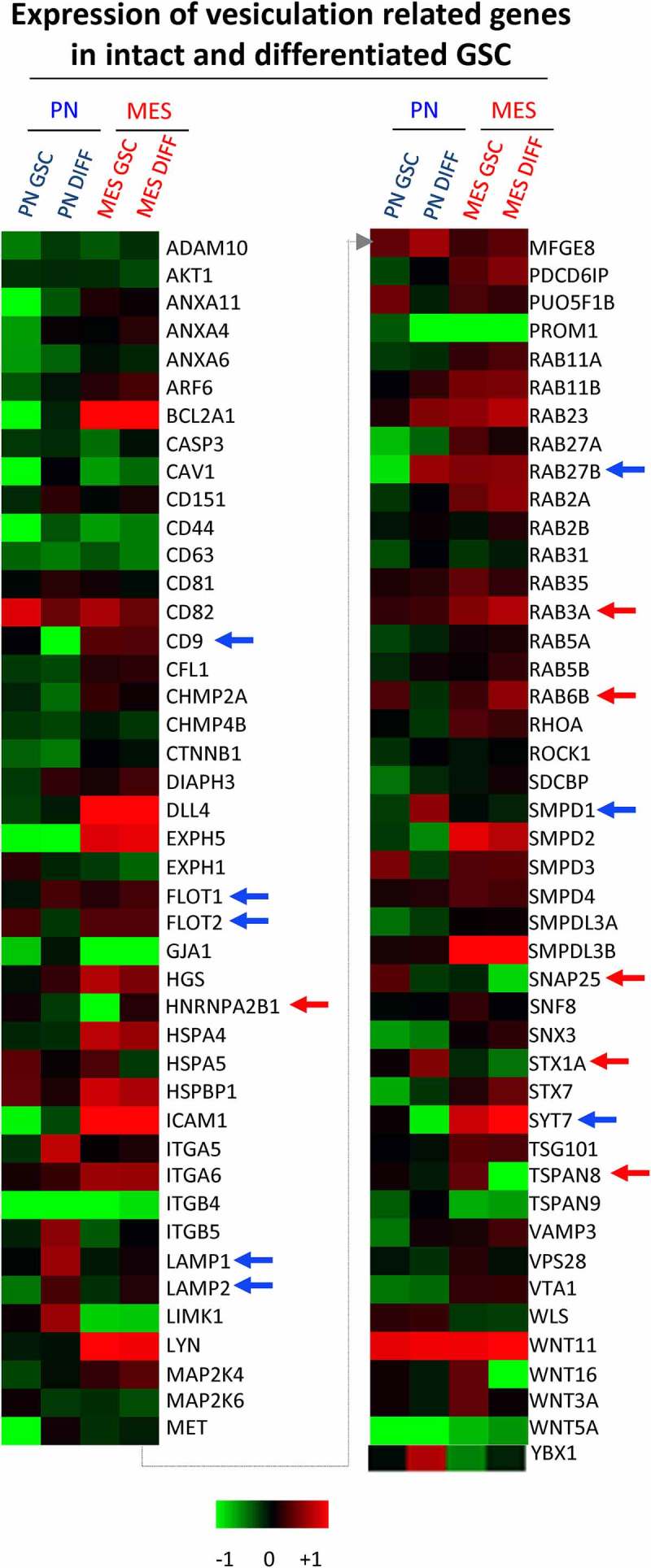
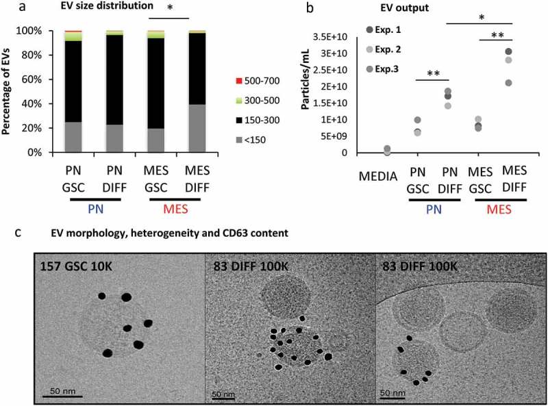
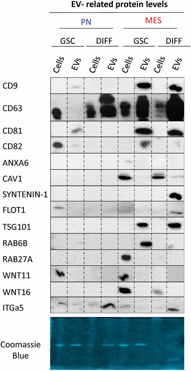
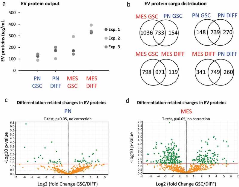
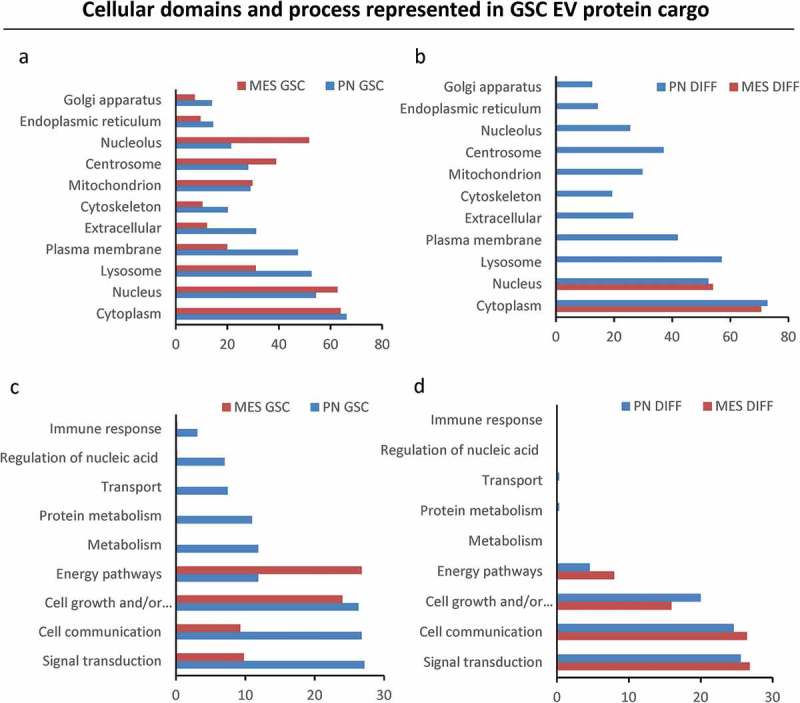
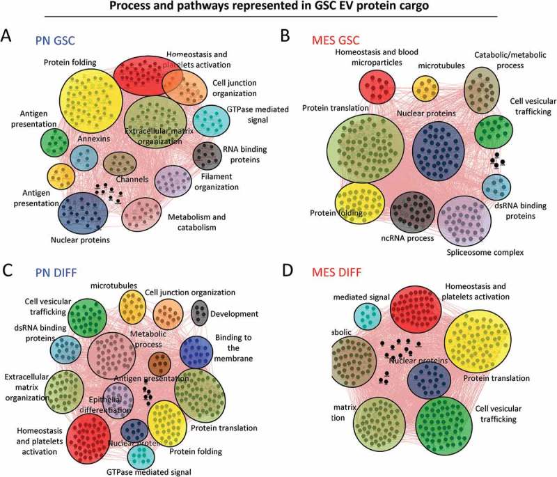
We have previously uncovered the impact of oncogenic and differentiation processes on extracellular vesicles (EVs) in cancer. This is of interested in the context of glioma stem cells (GSC) that are responsible for recurrent nature of glioblastoma multiforme (GBM), while retaining the potential to undergo differentiation and self renewal.  GSCs reside in vascular niches where they interact with endothelial cells through a number of mediators including bioactive cargo of EVs. GSCs can be classified as proneural (PN) or mesenchymal (MES) subtypes on the basis of their gene expression profiles and distinct biological characteristics. In the present study we investigated how GSC diversity and differentiation programmes influence their EV-mediated communication potentials. Indeed, molecular subtypes of GBMs and GSCs differ with respect to their expression of EV-related genes (vesiculome) and GSCs with PN or MES phenotypes produce EVs with markedly different characteristics, marker profiles, proteomes and endothelial stimulating activities. For example, while EVs of PN GSC are largely devoid of exosomal markers their counterparts from MES GSCs express ample CD9, CD63 and CD81 tetraspanins. In both GSC subtypes serum-induced differentiation results in profound, but distinct changes of cellular phenotypes including the enhanced EV production, reconfiguration of their proteomes and the related functional pathways. Notably, the EV uptake was a function of both subtype and differentiation state of donor cells. Thus, while, EVs produced by differentiated MES GSCs were internalized less efficiently than those from undifferentiated cells they exhibited an increased stimulatory potential for human brain endothelial cells. Such stimulating activity was also observed for EVs derived from differentiated PN GSCs, despite their even weaker uptake by endothelial cells. These findings suggest that the role of EVs as biological mediators and biomarkers in GBM may depend on the molecular subtype and functional state of donor cancer cells, including cancer stem cells. : CryoTEM: cryo-transmission electron microscopy; DIFF: differentiated GSCs; EGF: epidermal growth factor; DUC: differential ultracentrifugation; EV: extracellular vesicle; FGF: fibroblast growth factor; GBM: glioblastoma multiforme; GFAP: glial fibrillary acidic protein; GO: gene ontology; GSC: glioma stem cells; HBEC-5i: human brain endothelial cells; MES: mesenchymal cells; MTS - [3-(4,5-dimethylthiazol-2-yl)-5-(3-carboxymethoxyphenyl)-2-(4-sulfophenyl)-2H-tetrazolium, inner salt; PMT1: proneural-to-mesenchyman transition cell line 1; PN: proneural cells; TEM: transmission electron microscopy; WB: western blotting.

摘要

我们之前已经揭示了致癌和分化过程对癌症细胞外囊泡(EVs)的影响。这在胶质母细胞瘤干细胞(GSC)的背景下很有意思,胶质母细胞瘤干细胞是多形性胶质母细胞瘤(GBM)复发的原因,同时保留了分化和自我更新的潜力。GSCs存在于血管微环境中,在那里它们通过多种介质与内皮细胞相互作用,包括EVs的生物活性货物。基于其基因表达谱和独特的生物学特征,GSCs可分为神经前体细胞(PN)或间充质细胞(MES)亚型。在本研究中,我们调查了GSC多样性和分化程序如何影响其EV介导的通讯潜力。事实上,GBM和GSCs的分子亚型在其EV相关基因(囊泡组)的表达方面存在差异,具有PN或MES表型的GSCs产生的EVs具有明显不同的特征、标志物谱、蛋白质组和内皮刺激活性。例如,虽然PN GSC的EVs在很大程度上缺乏外泌体标志物,但MES GSCs的EVs表达丰富的CD9、CD63和CD81四跨膜蛋白。在两种GSC亚型中,血清诱导的分化都会导致细胞表型发生深刻但不同的变化,包括EV产生增加、蛋白质组重新配置以及相关功能途径。值得注意的是,EV摄取是供体细胞亚型和分化状态的函数。因此,虽然分化的MES GSCs产生的EVs比未分化细胞产生的EVs内化效率更低,但它们对人脑血管内皮细胞的刺激潜力增加。对于来自分化的PN GSCs的EVs也观察到了这种刺激活性,尽管它们被内皮细胞摄取的能力更弱。这些发现表明,EVs作为GBM中的生物介质和生物标志物的作用可能取决于供体癌细胞,包括癌症干细胞的分子亚型和功能状态。:冷冻透射电子显微镜;DIFF:分化的GSCs;表皮生长因子;差速超速离心;EV:细胞外囊泡;成纤维细胞生长因子;GBM:多形性胶质母细胞瘤;胶质纤维酸性蛋白;基因本体论;GSC:胶质母细胞瘤干细胞;人脑血管内皮细胞;MES:间充质细胞;MTS - [3 - (4,5 - 二甲基噻唑 - 2 - 基) - 5 - (3 - 羧甲氧基苯基) - 2 - (4 - 磺基苯基) - 2H - 四唑,内盐;PMT1:神经前体细胞向间充质细胞转变细胞系1;PN:神经前体细胞;透射电子显微镜;蛋白质免疫印迹

https://cdn.ncbi.nlm.nih.gov/pmc/blobs/c65f/6052423/c52502b0c056/ZJEV_A_1490144_F0010_OC.jpg
https://cdn.ncbi.nlm.nih.gov/pmc/blobs/c65f/6052423/a177a47ffa7f/ZJEV_A_1490144_F0001_OC.jpg
https://cdn.ncbi.nlm.nih.gov/pmc/blobs/c65f/6052423/cc1d310ba3f8/ZJEV_A_1490144_F0002_OC.jpg
https://cdn.ncbi.nlm.nih.gov/pmc/blobs/c65f/6052423/dc239213b007/ZJEV_A_1490144_F0003_OC.jpg
https://cdn.ncbi.nlm.nih.gov/pmc/blobs/c65f/6052423/37974c803b7f/ZJEV_A_1490144_F0004_OC.jpg
https://cdn.ncbi.nlm.nih.gov/pmc/blobs/c65f/6052423/925f9bb94f34/ZJEV_A_1490144_F0005_OC.jpg
https://cdn.ncbi.nlm.nih.gov/pmc/blobs/c65f/6052423/df8b6a2ebe04/ZJEV_A_1490144_F0006_OC.jpg
https://cdn.ncbi.nlm.nih.gov/pmc/blobs/c65f/6052423/2ff09c88b92b/ZJEV_A_1490144_F0007_OC.jpg
https://cdn.ncbi.nlm.nih.gov/pmc/blobs/c65f/6052423/0466ef4a8677/ZJEV_A_1490144_F0008_OC.jpg
https://cdn.ncbi.nlm.nih.gov/pmc/blobs/c65f/6052423/0edb00204e38/ZJEV_A_1490144_F0009_OC.jpg
https://cdn.ncbi.nlm.nih.gov/pmc/blobs/c65f/6052423/c52502b0c056/ZJEV_A_1490144_F0010_OC.jpg

相似文献

1
Molecular subtypes and differentiation programmes of glioma stem cells as determinants of extracellular vesicle profiles and endothelial cell-stimulating activities.胶质瘤干细胞的分子亚型和分化程序作为细胞外囊泡谱和内皮细胞刺激活性的决定因素
J Extracell Vesicles. 2018 Jul 17;7(1):1490144. doi: 10.1080/20013078.2018.1490144. eCollection 2018.
2
Gene expression profiling distinguishes proneural glioma stem cells from mesenchymal glioma stem cells.基因表达谱分析可区分神经干细胞样胶质瘤干细胞与间充质胶质瘤干细胞。
Genom Data. 2015 Sep 1;5:333-336. doi: 10.1016/j.gdata.2015.07.007.
3
Stem cell signature in glioblastoma: therapeutic development for a moving target.胶质母细胞瘤中的干细胞特征:针对动态靶点的治疗进展
J Neurosurg. 2015 Feb;122(2):324-30. doi: 10.3171/2014.9.JNS132253. Epub 2014 Nov 14.
4
Mesenchymal glioma stem cells are maintained by activated glycolytic metabolism involving aldehyde dehydrogenase 1A3.间质神经胶质瘤干细胞由激活的糖酵解代谢维持,该代谢涉及醛脱氢酶 1A3。
Proc Natl Acad Sci U S A. 2013 May 21;110(21):8644-9. doi: 10.1073/pnas.1221478110. Epub 2013 May 6.
5
Extracellular vesicles in the biology of brain tumour stem cells--Implications for inter-cellular communication, therapy and biomarker development.脑肿瘤干细胞生物学中的细胞外囊泡——对细胞间通讯、治疗和生物标志物开发的启示。
Semin Cell Dev Biol. 2015 Apr;40:17-26. doi: 10.1016/j.semcdb.2015.02.011. Epub 2015 Feb 23.
6
Extracellular vesicles produced by irradiated endothelial or Glioblastoma stem cells promote tumor growth and vascularization modulating tumor microenvironment.由受辐照的内皮细胞或胶质母细胞瘤干细胞产生的细胞外囊泡通过调节肿瘤微环境促进肿瘤生长和血管生成。
Cancer Cell Int. 2024 Feb 12;24(1):72. doi: 10.1186/s12935-024-03253-0.
7
The Impact of Oncogenic EGFRvIII on the Proteome of Extracellular Vesicles Released from Glioblastoma Cells.致癌性 EGFRvIII 对胶质母细胞瘤细胞释放的细胞外囊泡蛋白质组的影响。
Mol Cell Proteomics. 2018 Oct;17(10):1948-1964. doi: 10.1074/mcp.RA118.000644. Epub 2018 Jul 13.
8
Divergent evolution of temozolomide resistance in glioblastoma stem cells is reflected in extracellular vesicles and coupled with radiosensitization.胶质母细胞瘤干细胞中替莫唑胺耐药的差异进化反映在细胞外囊泡中,并与放射增敏相关。
Neuro Oncol. 2018 Jan 22;20(2):236-248. doi: 10.1093/neuonc/nox142.
9
Glioma-Derived miRNA-Containing Extracellular Vesicles Induce Angiogenesis by Reprogramming Brain Endothelial Cells.脑胶质瘤来源的含有 miRNA 的细胞外囊泡通过重编程脑内皮细胞诱导血管生成。
Cell Rep. 2020 Feb 18;30(7):2065-2074.e4. doi: 10.1016/j.celrep.2020.01.073.
10
Glioblastoma stem-like cells secrete the pro-angiogenic VEGF-A factor in extracellular vesicles.胶质母细胞瘤干细胞样细胞在细胞外囊泡中分泌促血管生成的血管内皮生长因子A(VEGF-A)。
J Extracell Vesicles. 2017 Aug 8;6(1):1359479. doi: 10.1080/20013078.2017.1359479. eCollection 2017.

引用本文的文献

1
Phenotypic variations in glioma stem cells: regulatory mechanisms and implications for therapeutic strategies.胶质瘤干细胞的表型变异:调控机制及其对治疗策略的影响
J Transl Med. 2025 Sep 2;23(1):984. doi: 10.1186/s12967-025-07034-9.
2
Glioblastoma: Overview of Proteomic Investigations and Biobank Approaches for the Development of a Multidisciplinary Translational Network.胶质母细胞瘤:蛋白质组学研究及生物样本库方法概述,用于构建多学科转化网络
Cancers (Basel). 2025 Jun 26;17(13):2151. doi: 10.3390/cancers17132151.
3
The Role of Extracellular Vesicles in the Control of Vascular Checkpoints for Cancer Metastasis.

本文引用的文献

1
Exosomes from Glioma-Associated Mesenchymal Stem Cells Increase the Tumorigenicity of Glioma Stem-like Cells via Transfer of miR-1587.来自胶质瘤相关间充质干细胞的外泌体通过miR-1587的传递增加胶质瘤干细胞样细胞的致瘤性。
Cancer Res. 2017 Nov 1;77(21):5808-5819. doi: 10.1158/0008-5472.CAN-16-2524. Epub 2017 Aug 30.
2
Fate mapping of human glioblastoma reveals an invariant stem cell hierarchy.人类胶质母细胞瘤的命运图谱揭示了一种不变的干细胞层级结构。
Nature. 2017 Sep 14;549(7671):227-232. doi: 10.1038/nature23666. Epub 2017 Aug 30.
3
Glioblastoma stem-like cells secrete the pro-angiogenic VEGF-A factor in extracellular vesicles.
细胞外囊泡在癌症转移血管检查点控制中的作用
Cancers (Basel). 2025 Jun 12;17(12):1966. doi: 10.3390/cancers17121966.
4
Exosomal miR-21-5p from glioma associated mesenchymal stem cells promotes the progression and glycolysis of glioblastoma via PDHA1.来自胶质瘤相关间充质干细胞的外泌体miR-21-5p通过PDHA1促进胶质母细胞瘤的进展和糖酵解。
Sci Rep. 2025 Jan 17;15(1):2320. doi: 10.1038/s41598-025-86580-0.
5
Prospects and Current Challenges of Extracellular Vesicle-Based Biomarkers in Cancer.基于细胞外囊泡的生物标志物在癌症中的前景与当前挑战
Biology (Basel). 2024 Sep 4;13(9):694. doi: 10.3390/biology13090694.
6
Extracellular vesicles in glioblastoma: a challenge and an opportunity.胶质母细胞瘤中的细胞外囊泡:挑战与机遇
NPJ Precis Oncol. 2024 May 17;8(1):103. doi: 10.1038/s41698-024-00600-2.
7
Comprehensive understanding of glioblastoma molecular phenotypes: classification, characteristics, and transition.全面了解胶质母细胞瘤的分子表型:分类、特征和转化。
Cancer Biol Med. 2024 May 6;21(5):363-81. doi: 10.20892/j.issn.2095-3941.2023.0510.
8
Mesenchymal glioma stem cells trigger vasectasia-distinct neovascularization process stimulated by extracellular vesicles carrying EGFR.间质神经胶质瘤干细胞通过携带 EGFR 的细胞外囊泡触发血管扩张——一种独特的血管新生过程。
Nat Commun. 2024 Apr 3;15(1):2865. doi: 10.1038/s41467-024-46597-x.
9
Proteomic, Metabolomic, and Fatty Acid Profiling of Small Extracellular Vesicles from Glioblastoma Stem-Like Cells and Their Role in Tumor Heterogeneity.脑胶质瘤干细胞来源的小细胞外囊泡的蛋白质组学、代谢组学和脂肪酸分析及其在肿瘤异质性中的作用。
ACS Nano. 2024 Jan 23;18(3):2500-2519. doi: 10.1021/acsnano.3c11427. Epub 2024 Jan 11.
10
Unlocking Glioblastoma Secrets: Natural Killer Cell Therapy against Cancer Stem Cells.揭开胶质母细胞瘤的秘密:自然杀伤细胞疗法对抗癌症干细胞。
Cancers (Basel). 2023 Dec 14;15(24):5836. doi: 10.3390/cancers15245836.
胶质母细胞瘤干细胞样细胞在细胞外囊泡中分泌促血管生成的血管内皮生长因子A(VEGF-A)。
J Extracell Vesicles. 2017 Aug 8;6(1):1359479. doi: 10.1080/20013078.2017.1359479. eCollection 2017.
4
Extracellular vesicle communication pathways as regulatory targets of oncogenic transformation.细胞外囊泡通讯途径作为致癌转化的调控靶点。
Semin Cell Dev Biol. 2017 Jul;67:11-22. doi: 10.1016/j.semcdb.2017.01.003. Epub 2017 Jan 8.
5
Epigenetic Activation of WNT5A Drives Glioblastoma Stem Cell Differentiation and Invasive Growth.WNT5A的表观遗传激活驱动胶质母细胞瘤干细胞分化和侵袭性生长。
Cell. 2016 Nov 17;167(5):1281-1295.e18. doi: 10.1016/j.cell.2016.10.039.
6
Barriers to horizontal cell transformation by extracellular vesicles containing oncogenic H-ras.含致癌性H-ras的细胞外囊泡介导水平细胞转化的障碍
Oncotarget. 2016 Aug 9;7(32):51991-52002. doi: 10.18632/oncotarget.10627.
7
Clonal evolution of glioblastoma under therapy.胶质母细胞瘤在治疗过程中的克隆进化。
Nat Genet. 2016 Jul;48(7):768-76. doi: 10.1038/ng.3590. Epub 2016 Jun 6.
8
Glioma.胶质瘤。
Nat Rev Dis Primers. 2015 Jul 16;1:15017. doi: 10.1038/nrdp.2015.17.
9
Extracellular vesicles and intercellular communication within the nervous system.细胞外囊泡与神经系统内的细胞间通讯。
J Clin Invest. 2016 Apr 1;126(4):1198-207. doi: 10.1172/JCI81134.
10
Extracellular Vesicles from High-Grade Glioma Exchange Diverse Pro-oncogenic Signals That Maintain Intratumoral Heterogeneity.高级别胶质瘤来源的细胞外囊泡交换多种促癌信号,维持肿瘤内异质性。
Cancer Res. 2016 May 15;76(10):2876-81. doi: 10.1158/0008-5472.CAN-15-3432. Epub 2016 Mar 24.