Laureate Institute for Brain Research, Tulsa, OK, United States.
Laureate Institute for Brain Research, Tulsa, OK, United States; Laureate Psychiatric Clinic and Hospital, Tulsa, OK, United States.
Neuroimage Clin. 2018 Apr 8;19:106-121. doi: 10.1016/j.nicl.2018.04.010. eCollection 2018.
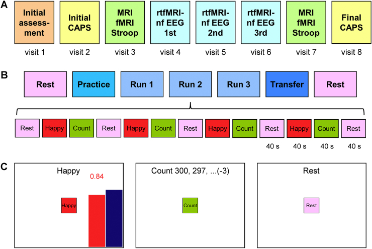
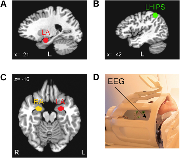
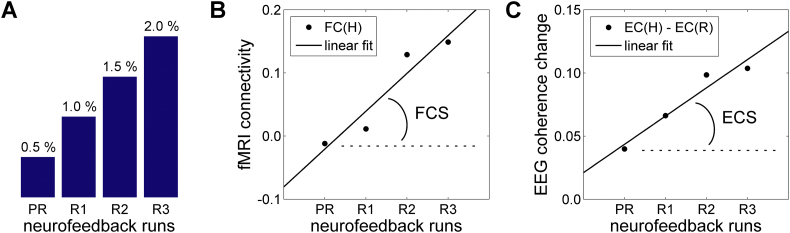
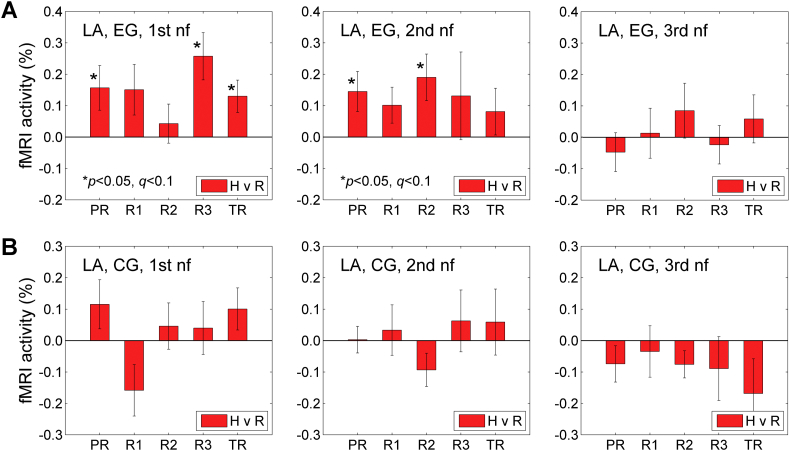
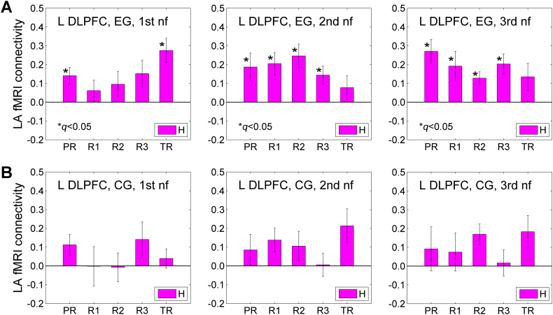
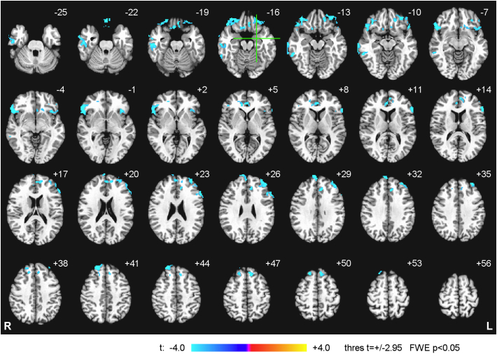
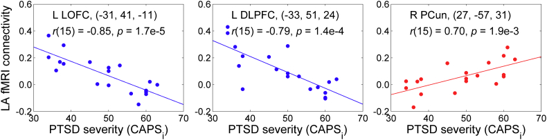
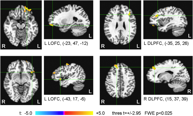
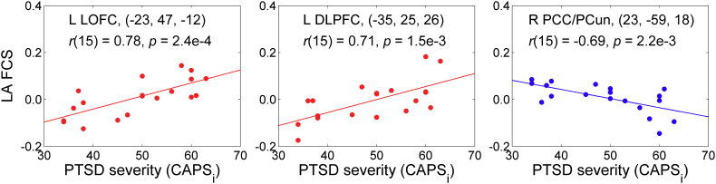
Posttraumatic stress disorder (PTSD) is a chronic and disabling neuropsychiatric disorder characterized by insufficient top-down modulation of the amygdala activity by the prefrontal cortex. Real-time fMRI neurofeedback (rtfMRI-nf) is an emerging method with potential for modifying the amygdala-prefrontal interactions. We report the first controlled emotion self-regulation study in veterans with combat-related PTSD utilizing rtfMRI-nf of the amygdala activity. PTSD patients in the experimental group (EG, = 20) learned to upregulate blood‑oxygenation-level-dependent (BOLD) activity of the left amygdala (LA) using the rtfMRI-nf during a happy emotion induction task. PTSD patients in the control group (CG, = 11) were provided with a sham rtfMRI-nf. The study included three rtfMRI-nf training sessions, and EEG recordings were performed simultaneously with fMRI. PTSD severity was assessed before and after the training using the Clinician-Administered PTSD Scale (CAPS). The EG participants who completed the study showed a significant reduction in total CAPS ratings, including significant reductions in avoidance and hyperarousal symptoms. They also exhibited a significant reduction in comorbid depression severity. Overall, 80% of the EG participants demonstrated clinically meaningful reductions in CAPS ratings, compared to 38% in the CG. No significant difference in the CAPS rating changes was observed between the groups. During the first rtfMRI-nf session, functional connectivity of the LA with the orbitofrontal cortex (OFC) and the dorsolateral prefrontal cortex (DLPFC) was progressively enhanced, and this enhancement significantly and positively correlated with the initial CAPS ratings. Left-lateralized enhancement in upper alpha EEG coherence also exhibited a significant positive correlation with the initial CAPS. Reduction in PTSD severity between the first and last rtfMRI-nf sessions significantly correlated with enhancement in functional connectivity between the LA and the left DLPFC. Our results demonstrate that the rtfMRI-nf of the amygdala activity has the potential to correct the amygdala-prefrontal functional connectivity deficiencies specific to PTSD.
创伤后应激障碍(PTSD)是一种慢性、致残性的神经精神疾病,其特征是前额叶皮层对杏仁核活动的自上而下调节不足。实时功能磁共振成像神经反馈(rtfMRI-nf)是一种新兴的方法,具有改变杏仁核-前额叶相互作用的潜力。我们报告了第一项利用 rtfMRI-nf 对与战斗相关 PTSD 的退伍军人进行的情绪自我调节的对照研究。实验组(EG,n=20)的 PTSD 患者在进行快乐情绪诱导任务时,学会通过 rtfMRI-nf 调节左杏仁核(LA)的血氧水平依赖(BOLD)活动。对照组(CG,n=11)的 PTSD 患者接受假 rtfMRI-nf。研究包括三个 rtfMRI-nf 训练疗程,同时进行 fMRI 和 EEG 记录。在训练前后使用临床医生管理的 PTSD 量表(CAPS)评估 PTSD 严重程度。完成研究的 EG 参与者的总 CAPS 评分显著降低,包括回避和警觉症状的显著降低。他们的共病抑郁严重程度也显著降低。总体而言,80%的 EG 参与者的 CAPS 评分有临床意义的降低,而 CG 组为 38%。两组 CAPS 评分变化无显著差异。在第一次 rtfMRI-nf 疗程中,LA 与眶额皮层(OFC)和背外侧前额叶皮层(DLPFC)的功能连接逐渐增强,这种增强与初始 CAPS 评分显著正相关。左偏侧化的上 alpha EEG 相干性增强也与初始 CAPS 呈显著正相关。第一次和最后一次 rtfMRI-nf 疗程之间 PTSD 严重程度的降低与 LA 和左 DLPFC 之间功能连接的增强显著相关。我们的结果表明,调节杏仁核活动的 rtfMRI-nf 有可能纠正 PTSD 特有的杏仁核-前额叶功能连接不足。