Brugarolas Pedro, Reich Daniel S, Popko Brian
1 Gordon Center for Medical Imaging, Massachusetts General Hospital and Harvard Medical School, Boston, MA, USA.
2 Translational Neuroradiology Section, National Institute of Neurological Disorders and Stroke, National Institutes of Health, Bethesda, MD, USA.
Mol Imaging. 2018 Jan-Dec;17:1536012118785471. doi: 10.1177/1536012118785471.
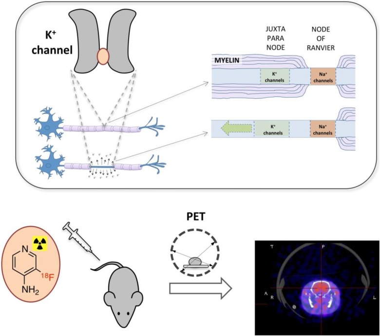
Noninvasive imaging of demyelination and remyelination is critical for diagnosis and clinical management of demyelinating diseases. Positron emission tomography (PET) has the potential to complement magnetic resonance imaging (MRI) by providing a quantitative measure specific to demyelination. In Brugarolas et al's study , we describe the development of the first PET tracer for voltage-gated K channels based on a clinically approved drug for multiple sclerosis that can be used for imaging demyelination in animal models.
脱髓鞘和再髓鞘化的无创成像对于脱髓鞘疾病的诊断和临床管理至关重要。正电子发射断层扫描(PET)有潜力通过提供针对脱髓鞘的定量测量来补充磁共振成像(MRI)。在布鲁加罗拉等人的研究中,我们描述了第一种基于一种临床上已批准用于治疗多发性硬化症的药物开发的用于电压门控钾通道的PET示踪剂,该示踪剂可用于动物模型中的脱髓鞘成像。