Vetrugno Luigi, Guadagnin Giovanni Maria, Orso Daniele, Boero Enrico, Bignami Elena, Bove Tiziana
Anesthesiology and Intensive Care Clinic, Department of Medicine, University of Udine, P.le S. Maria della Misericordia n.15, 33100, Udine, Italy.
Anesthesiology and Intensive Care, Department of Surgical Sciences, University of Turin, Turin, Italy.
Crit Ultrasound J. 2018 Aug 1;10(1):18. doi: 10.1186/s13089-018-0098-z.
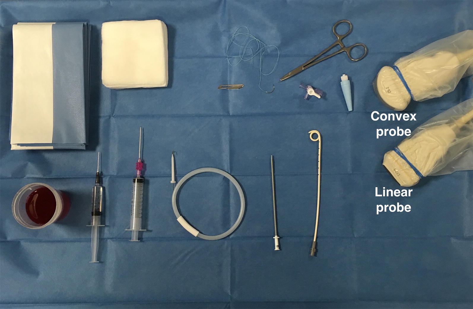
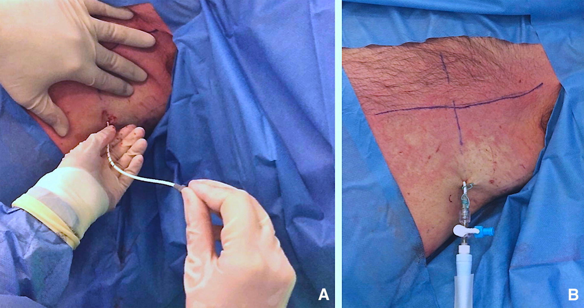
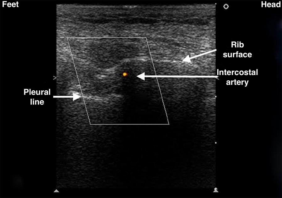
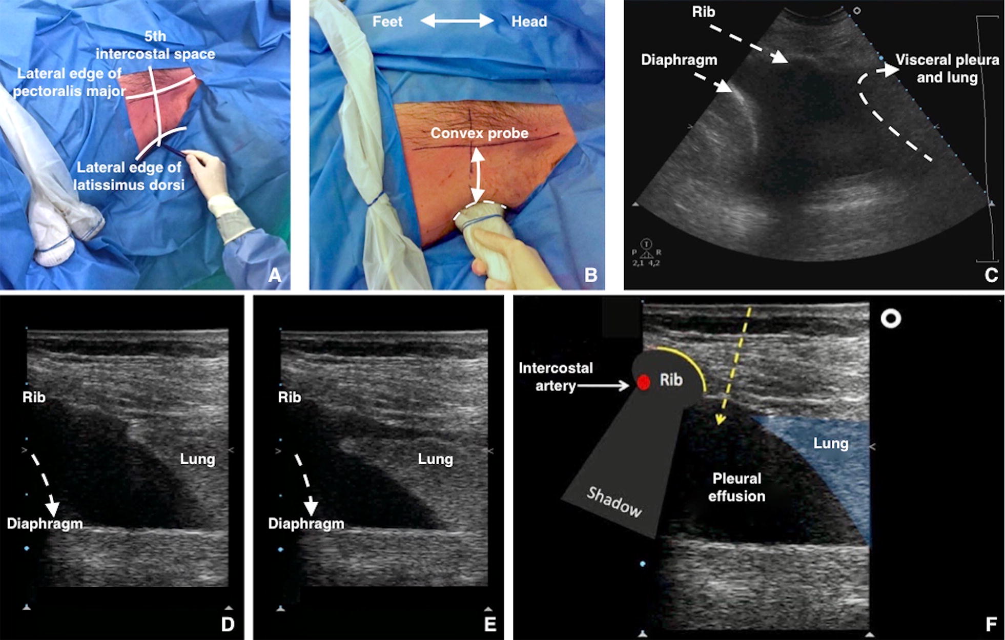
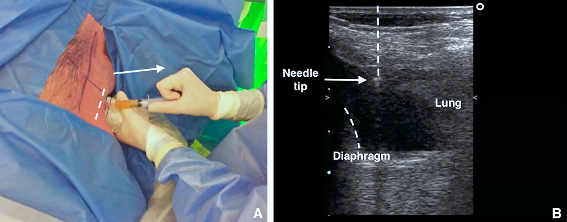
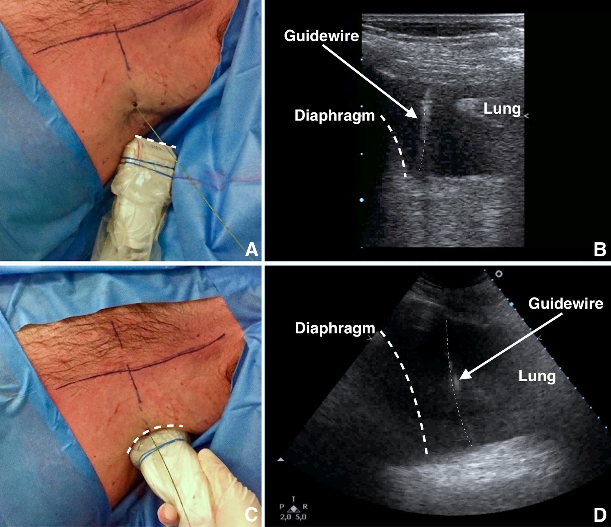
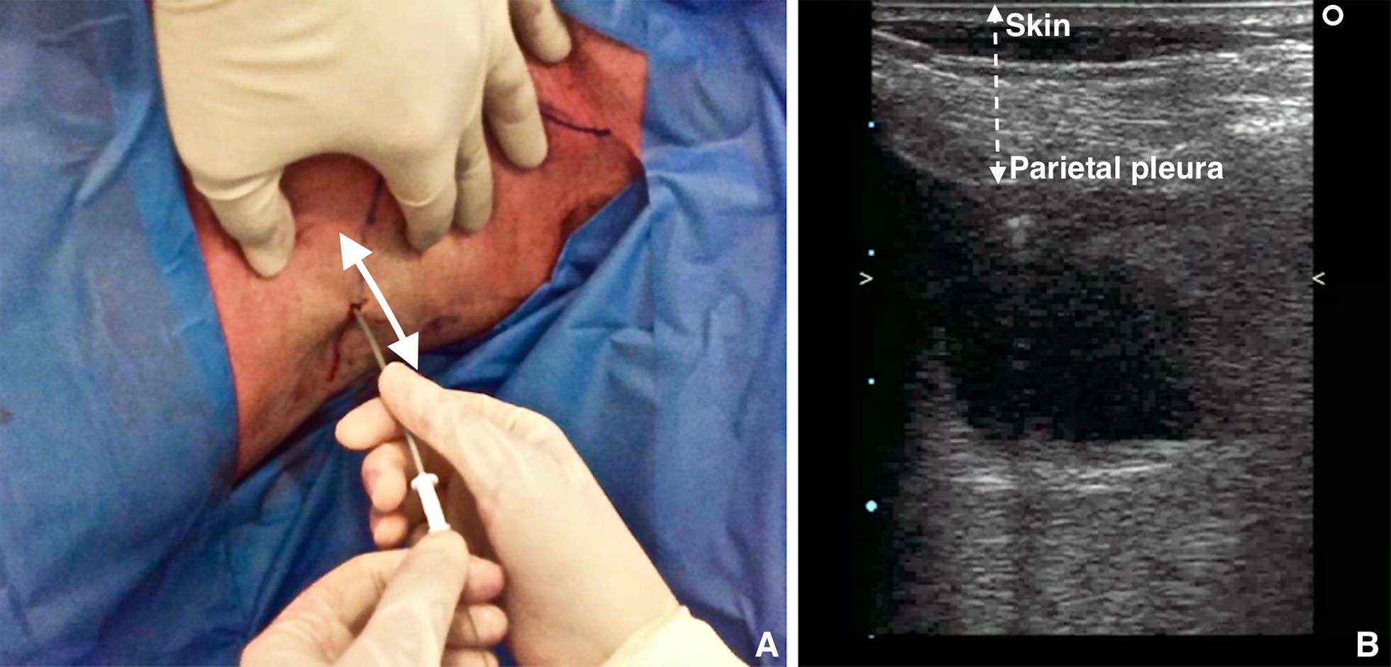
Thoracic ultrasound is a powerful diagnostic imaging technique for pleural space disorders. In addition to visualising pleural effusion, thoracic ultrasound also helps clinicians to identify the best puncture site and to guide the drainage insertion procedure. Thoracic ultrasound is essential during these invasive manoeuvres to increase safety and decrease potential life-threatening complications. This paper provides a technical description of pigtail-type drainage insertion using thoracic ultrasound, paying particular attention to indications, contraindications, ultrasound guidance, preparation/equipment, procedure and complications.
胸部超声是用于诊断胸膜腔疾病的一种强大的成像技术。除了可显示胸腔积液外,胸部超声还能帮助临床医生确定最佳穿刺部位并指导引流管置入操作。在这些侵入性操作过程中,胸部超声对于提高安全性和减少潜在的危及生命的并发症至关重要。本文提供了使用胸部超声进行猪尾型引流管置入的技术描述,特别关注适应证、禁忌证、超声引导、准备/设备、操作步骤及并发症。