Department of Medicine 1, University of Erlangen-Nuremberg, Erlangen, Germany.
Department of Pathology, University of Erlangen-Nuremberg, Erlangen, Germany.
Med Sci Monit. 2018 Aug 5;24:5437-5447. doi: 10.12659/MSM.909989.
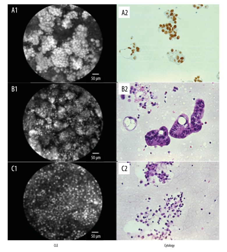
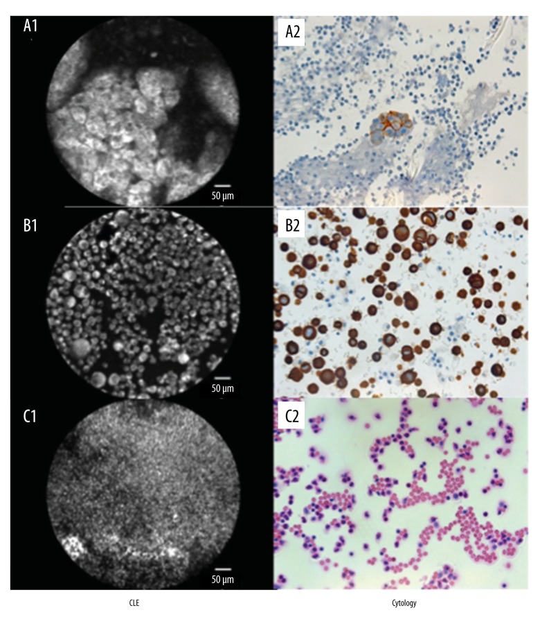
BACKGROUND Confocal laser endomicroscopy (CLE) enables "in vivo" microscopic tissue diagnosis based on tissue reflectance or tissue fluorescence upon application of fluorescence agents. The aim of the present study was to evaluate CLE as a new diagnostic approach for differentiation between malignant versus non-malignant pleural effusions. MATERIAL AND METHODS In 100 patients with pleural effusions, thoracentesis was performed. Cresyl violet and acriflavine were used as contrast agents for probe-based CLE of effusions. CLE video sequences were assessed by 4 independent investigators (2 experienced in this technique, 2 with only basic knowledge). In addition, all CLE samples were evaluated by an expert pathologist (p). Results were compared with conventional cytology of effusions and histology of cell blocks. RESULTS CLE reliably permitted identification of malignant cells in pleural effusions. Sensitivity for detection of malignant effusions was 87% (p: 87%) and 81% (p: 72%) for acriflavine and cresyl violet, respectively. With regard to specificity, acriflavine and cresyl violet yielded a mean value of 99% (p: 100%) and 92% (p: 100%). CONCLUSIONS In this pilot study, CLE permitted simple and rapid detection of malignant pleural effusions. Larger prospective studies are warranted to corroborate our findings.
共聚焦激光内镜检查(CLE)可通过应用荧光剂后对组织反射或组织荧光进行“体内”微观组织诊断。本研究旨在评估 CLE 作为一种新的诊断方法,用于区分恶性与非恶性胸腔积液。
在 100 例胸腔积液患者中进行了胸腔穿刺术。使用甲紫和吖啶橙作为基于探头的胸腔积液 CLE 的对比剂。由 4 位独立的研究人员(2 位有该技术经验,2 位仅具备基础知识)评估 CLE 视频序列。此外,所有 CLE 样本均由专家病理学家(p)评估。结果与常规细胞学和细胞块组织学进行比较。
CLE 可可靠地识别胸腔积液中的恶性细胞。吖啶橙和甲紫检测恶性胸腔积液的敏感性分别为 87%(p:87%)和 81%(p:72%)。关于特异性,吖啶橙和甲紫的平均值分别为 99%(p:100%)和 92%(p:100%)。
在这项初步研究中,CLE 可简单快速地检测恶性胸腔积液。需要更大的前瞻性研究来证实我们的发现。