Sasaki Akiko, Egashira Hideto, Sugimoto Hideyasu, Seki Kenichi, Tsukiyama Toshitaka, Ichita Chikamasa, Tokoro Shinnosuke, Takizawa Satoshi, Kitagawa Izumi, Teshima Shinichi, Kako Makoto
Gastroenterology Medicine Center, Shonan Kamakura General Hospital, Japan.
Pulmonology, Shonan Kamakura General Hospital, Japan.
Intern Med. 2018 Dec 15;57(24):3631-3635. doi: 10.2169/internalmedicine.1063-18. Epub 2018 Aug 10.
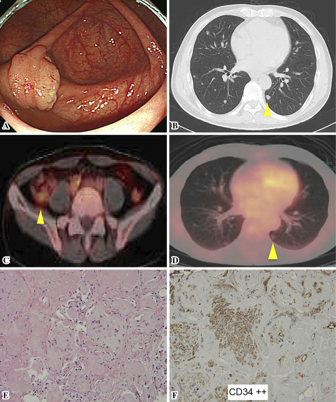
A 69-year-old male patient presented with multiple lung nodules revealed by chest-computed tomography (CT) during a preoperative examination for an appendiceal tumor. The nodule diameters ranged from 2-10 mm without either pleural thickening or effusions. A fluorine-18-labeled fluorodeoxyglucose (F-FDG)-positron emission tomography (PET)/CT scan showed a high FDG uptake in the appendiceal tumor, but almost normal standardized uptake values in the bilateral lung nodules. A CT-guided biopsy led to a diagnosis of pulmonary epithelioid hemangioendothelioma, a rare vascular tumor with a radiological presentation similar to that of a metastatic lung tumor. The present case is the first to describe successful treatment using a CT-guided biopsy instead of more conventional methods.
一名69岁男性患者在阑尾肿瘤术前检查时,胸部计算机断层扫描(CT)发现多个肺结节。结节直径为2 - 10毫米,无胸膜增厚或胸腔积液。氟-18标记的氟脱氧葡萄糖(F-FDG)正电子发射断层扫描(PET)/CT显示阑尾肿瘤有高FDG摄取,但双侧肺结节的标准化摄取值几乎正常。CT引导下活检诊断为肺上皮样血管内皮瘤,这是一种罕见的血管肿瘤,其影像学表现与肺转移瘤相似。本病例是首例描述使用CT引导活检而非更传统方法成功治疗的案例。