Department of Medicine, University of Massachusetts Medical School, Worcester, MA.
Institute and Department of Infectious Disease, Tongji Hospital, Tongji Medical College, Huazhong University of Science and Technology, Wuhan, China.
Hepatology. 2019 Feb;69(2):545-563. doi: 10.1002/hep.30215. Epub 2019 Jan 4.
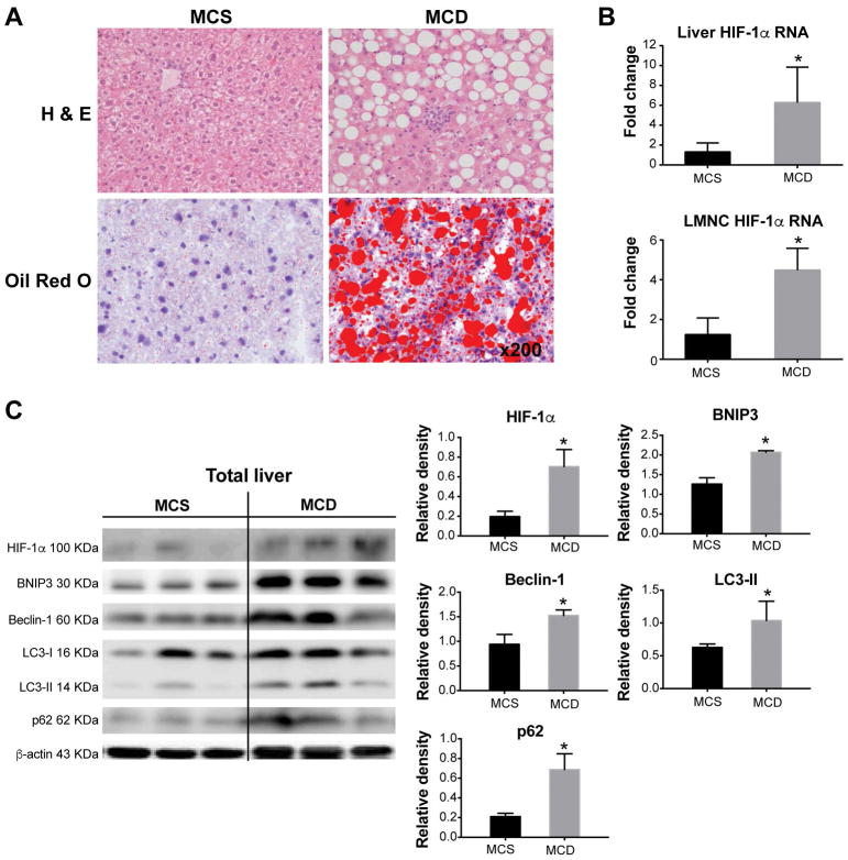
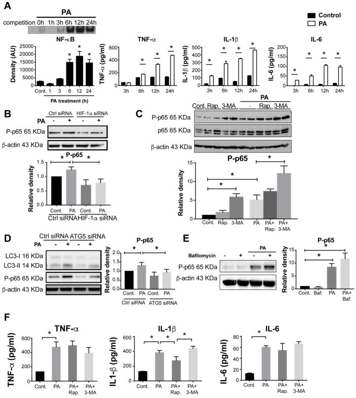
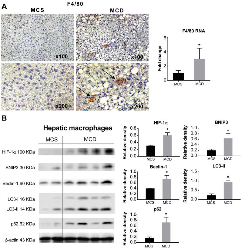
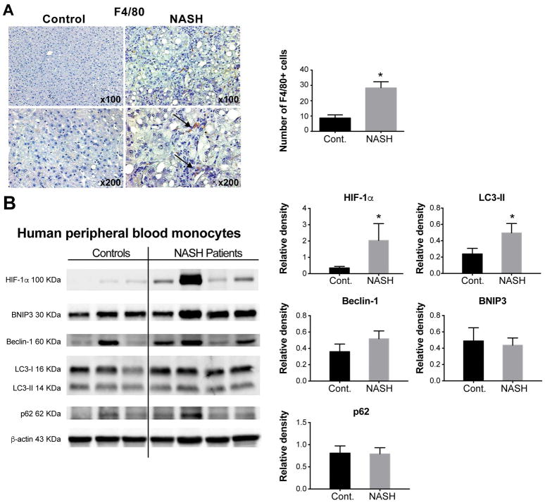
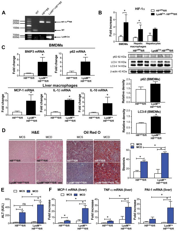
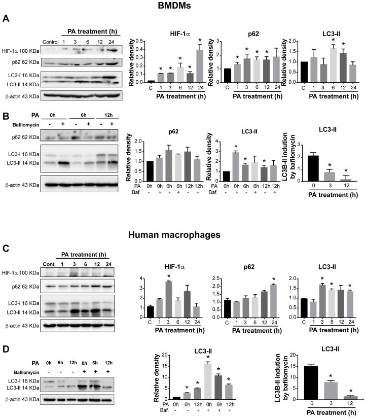
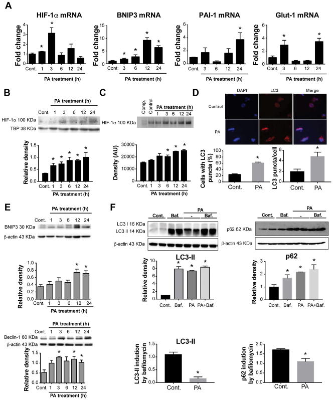
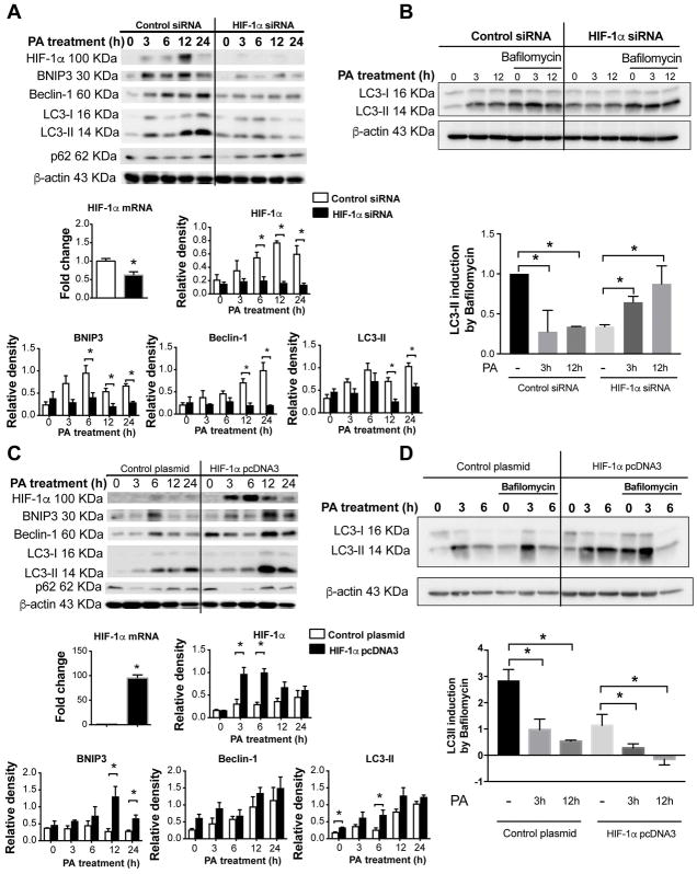
Inflammatory cell activation drives diverse cellular programming during hepatic diseases. Hypoxia-inducible factors (HIFs) have recently been identified as important regulators of immunity and inflammation. In nonalcoholic steatohepatitis (NASH), HIF-1α is upregulated in hepatocytes, where it induces steatosis; however, the role of HIF-1α in macrophages under metabolic stress has not been explored. In this study, we found increased HIF-1α levels in hepatic macrophages in methionine-choline-deficient (MCD) diet-fed mice and in macrophages of patients with NASH compared with controls. The HIF-1α increase was concomitant with elevated levels of autophagy markers BNIP3, Beclin-1, LC3-II, and p62 in both mouse and human macrophages. LysM HIF fl/fl mice, which have HIF-1α levels stabilized in macrophages, showed higher steatosis and liver inflammation compared with HIF fl/fl mice on MCD diet. In vitro and ex vivo experiments reveal that saturated fatty acid, palmitic acid (PA), both induces HIF-1α and impairs autophagic flux in macrophages. Using small interfering RNA-mediated knock-down and overexpression of HIF-1α in macrophages, we demonstrated that PA impairs autophagy via HIF-1α. We found that HIF-1α mediates NF-κB activation and MCP-1 production and that HIF-1α-mediated impairment of macrophage autophagy increases IL-1β production, contributing to MCD diet-induced NASH. Conclusion: Palmitic acid impairs autophagy via HIF-1α activation in macrophages. HIF-1α and impaired autophagy are present in NASH in vivo in mouse macrophages and in human blood monocytes. We identified that HIF-1α activation and decreased autophagic flux stimulate inflammation in macrophages through upregulation of NF-κB activation. These results suggest that macrophage activation in NASH involves a complex interplay between HIF-1α and autophagy as these pathways promote proinflammatory overactivation in MCD diet-induced NASH.
炎症细胞的激活驱动着肝脏疾病中多种细胞程序的发生。缺氧诱导因子(HIFs)最近被鉴定为免疫和炎症的重要调节因子。在非酒精性脂肪性肝炎(NASH)中,HIF-1α在肝细胞中上调,诱导脂肪变性;然而,在代谢应激下,HIF-1α在巨噬细胞中的作用尚未被探索。在这项研究中,我们发现,在蛋氨酸-胆碱缺乏(MCD)饮食喂养的小鼠和 NASH 患者的巨噬细胞中,HIF-1α 水平升高。在人和鼠巨噬细胞中,HIF-1α 的增加伴随着自噬标志物 BNIP3、Beclin-1、LC3-II 和 p62 水平的升高。LysM HIF fl/fl 小鼠中,巨噬细胞中的 HIF-1α 水平稳定,与 MCD 饮食喂养的 HIF fl/fl 小鼠相比,表现出更高的脂肪变性和肝脏炎症。体外和体内实验表明,饱和脂肪酸棕榈酸(PA)既诱导 HIF-1α 的表达,又损害巨噬细胞中的自噬通量。通过巨噬细胞中的小干扰 RNA 介导的敲低和 HIF-1α 的过表达,我们证明了 PA 通过 HIF-1α 损害自噬。我们发现 HIF-1α 介导 NF-κB 激活和 MCP-1 的产生,并且 HIF-1α 介导的巨噬细胞自噬受损增加了 IL-1β 的产生,导致 MCD 饮食诱导的 NASH。结论:棕榈酸通过激活 HIF-1α 损害巨噬细胞中的自噬。在 MCD 饮食诱导的 NASH 中,我们在体内的小鼠巨噬细胞和人类血液单核细胞中发现了 HIF-1α 和受损的自噬。我们发现,HIF-1α 的激活和自噬通量的降低通过上调 NF-κB 激活刺激巨噬细胞中的炎症。这些结果表明,NASH 中的巨噬细胞激活涉及 HIF-1α 和自噬之间的复杂相互作用,因为这些途径促进了 MCD 饮食诱导的 NASH 中促炎的过度激活。