Center for Quantitative Analysis of Molecular and Cellular Biosystems (BioQuant), Heidelberg University, Heidelberg, Germany.
Division of Theoretical Bioinformatics, German Cancer Research Center (DKFZ), Heidelberg, Germany.
Mol Syst Biol. 2018 Aug 13;14(8):e8238. doi: 10.15252/msb.20188238.
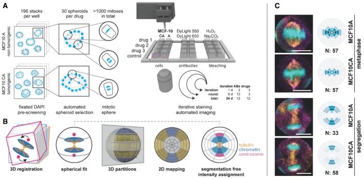
Three-dimensional protein localization intricately determines the functional coordination of cellular processes. The complex spatial context of protein landscape has been assessed by multiplexed immunofluorescent staining or mass spectrometry, applied to 2D cell culture with limited physiological relevance or tissue sections. Here, we present 3D SPECS, an automated technology for 3D Spatial characterization of Protein Expression Changes by microscopic Screening. This workflow comprises iterative antibody staining, high-content 3D imaging, and machine learning for detection of mitoses. This is followed by mapping of spatial protein localization into a spherical, cellular coordinate system, a basis for model-based prediction of spatially resolved affinities of proteins. As a proof-of-concept, we mapped twelve epitopes in 3D-cultured spheroids and investigated the network effects of twelve mitotic cancer drugs. Our approach reveals novel insights into spindle fragility and chromatin stress, and predicts unknown interactions between proteins in specific mitotic pathways. 3D SPECS's ability to map potential drug targets by multiplexed immunofluorescence in 3D cell culture combined with our automated high-content assay will inspire future functional protein expression and drug assays.
三维蛋白质定位精细地决定了细胞过程的功能协调。蛋白质景观的复杂空间背景已经通过多重免疫荧光染色或质谱分析来评估,这些方法应用于具有有限生理相关性的 2D 细胞培养或组织切片。在这里,我们提出了 3D SPECS,这是一种通过显微镜筛选对蛋白质表达变化进行三维空间特征描述的自动化技术。该工作流程包括迭代抗体染色、高内涵 3D 成像和机器学习,以检测有丝分裂。接着,将空间蛋白质定位映射到一个球形的细胞坐标系中,这是基于模型的蛋白质空间分辨率亲和力预测的基础。作为概念验证,我们在 3D 培养的球体中绘制了十二个表位,并研究了十二个有丝分裂癌症药物的网络效应。我们的方法揭示了纺锤体脆弱性和染色质应激的新见解,并预测了特定有丝分裂途径中蛋白质之间未知的相互作用。3D SPECS 能够在 3D 细胞培养中通过多重免疫荧光来绘制潜在的药物靶点,结合我们的自动化高内涵检测,将激发未来功能性蛋白质表达和药物检测的研究。