He Yujun, Wen Jin, Cui Qinghua, Lai Fangfang, Yin Dali, Cui Huaqing
State Key Laboratory of Bioactive Substance and Function of Natural Medicines, Institute of Materia Medica, Chinese Academy of Medical Sciences and Peking Union Medical College, Beijing, China.
Department of Urology, Peking Union Medical College Hospital, Beijing, China.
Front Pharmacol. 2018 Jul 25;9:812. doi: 10.3389/fphar.2018.00812. eCollection 2018.
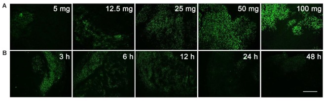
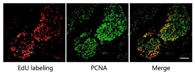
Current methods used to evaluate target efficacy of selected compound include western blot to semi-quantitatively analyze protein expression. However, problems arise as it is difficult to compare target efficacy of anti-tumor agents with the same mode of action. It is therefore desirable to develop a protocol that can quantitatively display target efficacy while also providing other useful information. In this study EdU labeling was used to mark out the proliferating area. The tumor tissue was accordingly divided into proliferating and non-proliferating areas. Fifteen tumor related proteins were stained by immunofluorescence and were found to express in either the proliferating or non-proliferating areas. This allows the quantitative analysis of protein expressions within the precise area. With simple image analysis, our method gave precise percent changes of protein expression and cell proliferation between the drugs treated group and the control group. Additional information, such as, the status of protein expression can also be obtained. This method exhibits high sensitivity, and provides a quantitative approach for evaluation of target efficacy.
目前用于评估所选化合物靶点疗效的方法包括蛋白质印迹法,用于半定量分析蛋白质表达。然而,由于难以比较具有相同作用模式的抗肿瘤药物的靶点疗效,因此出现了问题。因此,需要开发一种方案,既能定量显示靶点疗效,又能提供其他有用信息。在本研究中,使用EdU标记来标记增殖区域。肿瘤组织因此被分为增殖区和非增殖区。通过免疫荧光对15种肿瘤相关蛋白进行染色,发现它们在增殖区或非增殖区表达。这使得能够在精确区域内对蛋白质表达进行定量分析。通过简单的图像分析,我们的方法给出了药物治疗组和对照组之间蛋白质表达和细胞增殖的精确百分比变化。还可以获得其他信息,例如蛋白质表达状态。该方法具有高灵敏度,并为评估靶点疗效提供了一种定量方法。