• 文献检索
  • 文档翻译
  • 深度研究
  • 学术资讯
  • Suppr Zotero 插件Zotero 插件
  • 邀请有礼
  • 套餐&价格
  • 历史记录
应用&插件
Suppr Zotero 插件Zotero 插件浏览器插件Mac 客户端Windows 客户端微信小程序
定价
高级版会员购买积分包购买API积分包
服务
文献检索文档翻译深度研究API 文档MCP 服务
关于我们
关于 Suppr公司介绍联系我们用户协议隐私条款
关注我们

Suppr 超能文献

核心技术专利:CN118964589B侵权必究
粤ICP备2023148730 号-1Suppr @ 2026

文献检索

告别复杂PubMed语法,用中文像聊天一样搜索,搜遍4000万医学文献。AI智能推荐,让科研检索更轻松。

立即免费搜索

文件翻译

保留排版,准确专业,支持PDF/Word/PPT等文件格式,支持 12+语言互译。

免费翻译文档

深度研究

AI帮你快速写综述,25分钟生成高质量综述,智能提取关键信息,辅助科研写作。

立即免费体验

脉络膜上腔出血后行扫频源光学相干断层扫描:一例报告

Suprachoroidal hemorrhage followed by swept-source optical coherence tomography: a case report.

作者信息

Uramoto Kengo, Shimada Noriaki, Takahashi Hiroyuki, Murai Hideki, Shinohara Kosei, Ohno-Matsui Kyoko

机构信息

Department of Ophthalmology and Visual Science, Tokyo Medical and Dental University, 1-5-45 Yushima, Bunkyo-ku, Tokyo, 113-8519, Japan.

出版信息

BMC Ophthalmol. 2018 Aug 20;18(1):203. doi: 10.1186/s12886-018-0881-4.

DOI:10.1186/s12886-018-0881-4
PMID:30126397
原文链接:https://pmc.ncbi.nlm.nih.gov/articles/PMC6102832/
Abstract

BACKGROUND

To report a case of Suprachoroidal Hemorrhage followed by Swept-Source Optical Coherence Tomography.

CASE PRESENTATION

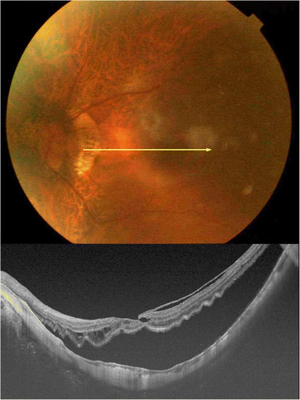
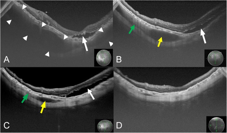
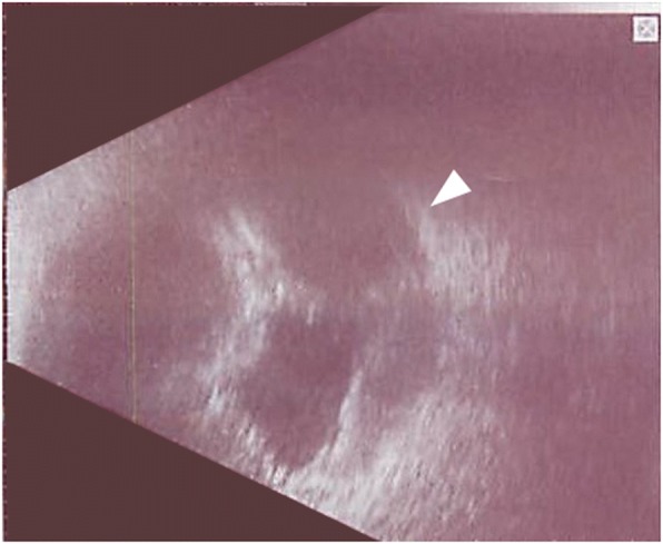
A 66-year-old woman with a rhegmatogenous retinal detachment in her left eye underwent pars plana vitrectomy. During the intraocular photocoagulation for a retinal tear after fluid-air exchange, a vitreous hemorrhage and suprachoroidal hemorrhage (SCH) developed. The surgical incisions were closed after filling the vitreous cavity with silicone oil. Two weeks later, the hemolyzed hemorrhage was removed, and new silicone oil was injected. After the surgery, a low reflective region was detected near the macula in the swept-source optical coherence tomographic (SS-OCT) images. The low reflective region was caused by the residual hemorrhage. The size of the reflective region gradually decreased and was not present at 3 months. We conclude that SS-OCT can be used to follow the resolution of a suprachoroidal hemorrhage.

CONCLUSION

SS-OCT can be used to detect and follow the natural course of a suprachoroidal hemorrhage including the absorptive processes.

摘要

背景

报告一例脉络膜上腔出血并随后进行扫频光学相干断层扫描的病例。

病例介绍

一名66岁左眼孔源性视网膜脱离的女性接受了玻璃体切除术。在液气交换后对视网膜裂孔进行眼内光凝时,发生了玻璃体出血和脉络膜上腔出血(SCH)。用硅油填充玻璃体腔后关闭手术切口。两周后,清除溶血的出血,并注入新的硅油。术后,在扫频光学相干断层扫描(SS-OCT)图像中黄斑附近检测到一个低反射区域。该低反射区域是由残留出血引起的。反射区域的大小逐渐减小,3个月时消失。我们得出结论,SS-OCT可用于跟踪脉络膜上腔出血的消退情况。

结论

SS-OCT可用于检测和跟踪脉络膜上腔出血的自然病程,包括吸收过程。

https://cdn.ncbi.nlm.nih.gov/pmc/blobs/4b57/6102832/d9892dc61a13/12886_2018_881_Fig3_HTML.jpg
https://cdn.ncbi.nlm.nih.gov/pmc/blobs/4b57/6102832/7f2d4fe56402/12886_2018_881_Fig1_HTML.jpg
https://cdn.ncbi.nlm.nih.gov/pmc/blobs/4b57/6102832/60cb65a56559/12886_2018_881_Fig2_HTML.jpg
https://cdn.ncbi.nlm.nih.gov/pmc/blobs/4b57/6102832/d9892dc61a13/12886_2018_881_Fig3_HTML.jpg
https://cdn.ncbi.nlm.nih.gov/pmc/blobs/4b57/6102832/7f2d4fe56402/12886_2018_881_Fig1_HTML.jpg
https://cdn.ncbi.nlm.nih.gov/pmc/blobs/4b57/6102832/60cb65a56559/12886_2018_881_Fig2_HTML.jpg
https://cdn.ncbi.nlm.nih.gov/pmc/blobs/4b57/6102832/d9892dc61a13/12886_2018_881_Fig3_HTML.jpg

相似文献

1
Suprachoroidal hemorrhage followed by swept-source optical coherence tomography: a case report.脉络膜上腔出血后行扫频源光学相干断层扫描:一例报告
BMC Ophthalmol. 2018 Aug 20;18(1):203. doi: 10.1186/s12886-018-0881-4.
2
Delayed Suprachoroidal Hemorrhage After Pars Plana Vitrectomy: Five-Year Results of a Retrospective Multicenter Cohort Study.玻璃体切割术后迟发性脉络膜上腔出血:一项回顾性多中心队列研究的五年结果
Am J Ophthalmol. 2015 Dec;160(6):1235-1242.e1. doi: 10.1016/j.ajo.2015.08.035. Epub 2015 Sep 4.
3
VISUALIZATION OF THE MACULA IN GAS-FILLED EYES: Spectral Domain Optical Coherence Tomography Versus Swept-Source Optical Coherence Tomography.充气体眼中黄斑的可视化:谱域光学相干断层扫描与扫频源光学相干断层扫描比较。
Retina. 2018 Mar;38(3):480-489. doi: 10.1097/IAE.0000000000001560.
4
Suprachoroidal hemorrhage associated with pars plana vitrectomy.与扁平部玻璃体切割术相关的脉络膜上腔出血。
BMC Ophthalmol. 2021 Aug 11;21(1):295. doi: 10.1186/s12886-021-02062-7.
5
Suprachoroidal buckling.脉络膜上腔扣带术
Dev Ophthalmol. 2014;54:135-46. doi: 10.1159/000360459. Epub 2014 Aug 26.
6
Optical Coherence Tomography Evaluation of Valsalva-Induced Suprachoroidal Hemorrhage.Valsalva动作诱发脉络膜上腔出血的光学相干断层扫描评估
Ophthalmic Surg Lasers Imaging Retina. 2016 Jul 1;47(7):674-6. doi: 10.3928/23258160-20160707-11.
7
When do you go back to the OR? A case of delayed IOL repositioning after suprachoroidal hemorrhage.
Can J Ophthalmol. 2019 Oct;54(5):e225-e227. doi: 10.1016/j.jcjo.2019.02.015. Epub 2019 Apr 9.
8
Swept-Source Optical Coherence Tomography Correlations Between Retina and Choroid Before and After Vitrectomy for Epiretinal Membranes.视网膜前膜玻璃体切除术前、后视网膜与脉络膜之间的扫频源光学相干断层扫描相关性研究
Am J Ophthalmol. 2016 May;165:100-7. doi: 10.1016/j.ajo.2016.02.003. Epub 2016 Mar 10.
9
Suprachoroidal hemorrhage during silicone oil removal in Marfan syndrome.马凡综合征患者硅油取出术中脉络膜上腔出血
Ophthalmic Surg Lasers. 2000 Jul-Aug;31(4):337-9.
10
Secondary surgical management of suprachoroidal hemorrhage during pars plana vitrectomy.玻璃体切除术中脉络膜上腔出血的二期手术处理
Vojnosanit Pregl. 2015 Mar;72(3):287-90. doi: 10.2298/vsp1503287v.

本文引用的文献

1
Minocycline attenuates photoreceptor degeneration in a mouse model of subretinal hemorrhage microglial: inhibition as a potential therapeutic strategy.米诺环素抑制视网膜下出血小鼠模型中小胶质细胞的光感受器变性:抑制作用作为一种潜在的治疗策略。
Am J Pathol. 2011 Sep;179(3):1265-77. doi: 10.1016/j.ajpath.2011.05.042. Epub 2011 Jul 19.
2
Suprachoroidal hemorrhage during pars plana vitrectomy.扁平部玻璃体切割术中脉络膜上腔出血
Curr Opin Ophthalmol. 2001 Jun;12(3):179-85. doi: 10.1097/00055735-200106000-00006.
3
Survey of risk factors for expulsive choroidal hemorrhage: case reports. Substantiation of the risk factors and their incidence.
驱逐性脉络膜出血危险因素的调查:病例报告。危险因素的证实及其发生率。
Ophthalmologica. 1996;210(6):344-7. doi: 10.1159/000310740.
4
Perioperative choroidal hemorrhage at pars plana vitrectomy. A case-control study.玻璃体切除术围手术期脉络膜出血。一项病例对照研究。
Ophthalmology. 1993 May;100(5):699-704. doi: 10.1016/s0161-6420(93)31586-1.
5
Expulsive hemorrhage: some observations and comments.驱逐性出血:一些观察与评论
Trans Am Ophthalmol Soc. 1974;72:157-69.