Gatto Rodolfo G, Amin Manish Y, Deyoung Daniel, Hey Matthew, Mareci Thomas H, Magin Richard L
1Department of Anatomy and Cell Biology, University of Illinois at Chicago, 808 S. Wood St. Rm 578 M/C 512, Chicago, IL 60612 USA.
2Department of Physics, University of Florida, Gainesville, FL USA.
Transl Neurodegener. 2018 Aug 8;7:20. doi: 10.1186/s40035-018-0122-z. eCollection 2018.
Amyotrophic lateral sclerosis (ALS) is a disease characterized by a progressive degeneration of motor neurons leading to paralysis. Our previous MRI diffusion tensor imaging studies detected early white matter changes in the spinal cords of mice carrying the G93A-SOD1 mutation. Here, we extend those studies using ultra-high field MRI (17.6 T) and fluorescent microscopy to investigate the appearance of early structural and connectivity changes in the spinal cords of ALS mice.
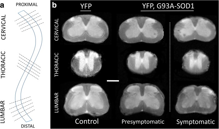
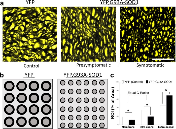
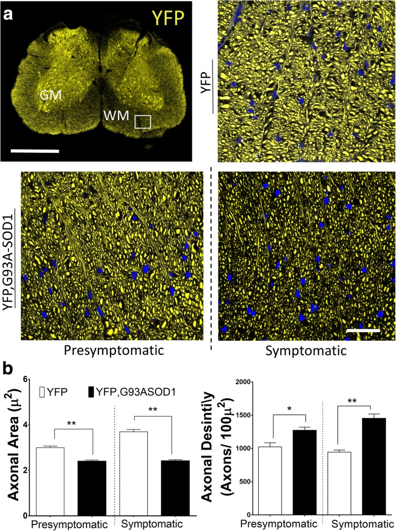
The spinal cords from presymptomatic and symptomatic mice (80 to 120 days of age) were scanned (ex-vivo) using diffusion-weighted MRI. The fractional anisotropy (FA), axial (AD) and radial (RD) diffusivities were calculated for axial slices from the thoracic, cervical and lumbar regions of the spinal cords. The diffusion parameters were compared with fluorescence microscopy and membrane cellular markers from the same tissue regions.
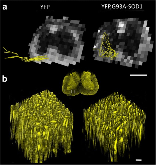
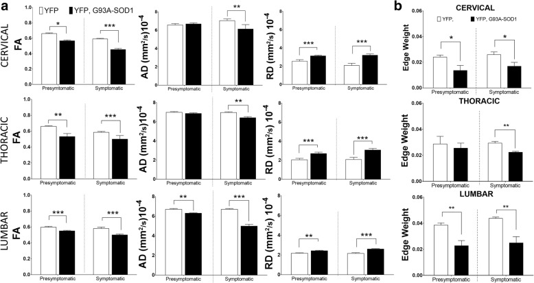
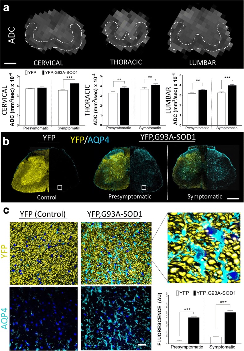
At early stages of the disease (day 80) in the lumbar region, we found, a 19% decrease in FA, a 9% decrease in AD and a 35% increase in RD. Similar changes were observed in cervical and thoracic spinal cord regions. Differences between control and ALS mice groups at the symptomatic stages (day 120) were larger. Quantitative fluorescence microscopy at 80 days, demonstrated a 22% reduction in axonal area and a 22% increase in axonal density. Tractography and quantitative connectome analyses measured by edge weights showed a 52% decrease in the lumbar regions of the spinal cords of this ALS mice group. A significant increase in ADC (23.3%) in the ALS mice group was related to an increase in aquaporin markers.
These findings suggest that the combination of ultra-high field diffusion MRI with fluorescent ALS mice reporters is a useful approach to detect and characterize presymptomatic white matter micro-ultrastructural changes and axonal connectivity anomalies in ALS.
肌萎缩侧索硬化症(ALS)是一种以运动神经元进行性退化导致瘫痪为特征的疾病。我们之前的磁共振成像扩散张量成像研究检测到携带G93A - SOD1突变的小鼠脊髓早期白质变化。在此,我们使用超高场磁共振成像(17.6 T)和荧光显微镜扩展这些研究,以调查ALS小鼠脊髓早期结构和连接性变化的表现。
对无症状和有症状小鼠(80至120日龄)的脊髓进行(离体)扩散加权磁共振成像扫描。计算脊髓胸段、颈段和腰段轴向切片的分数各向异性(FA)、轴向扩散率(AD)和径向扩散率(RD)。将扩散参数与来自相同组织区域的荧光显微镜和膜细胞标记物进行比较。
在疾病早期(第80天)的腰段区域,我们发现FA降低了19%,AD降低了9%,RD增加了35%。在颈段和胸段脊髓区域也观察到类似变化。有症状阶段(第120天)对照组和ALS小鼠组之间的差异更大。80天时的定量荧光显微镜检查显示轴突面积减少了22%,轴突密度增加了22%。通过边缘权重测量的纤维束成像和定量连接组分析显示,该ALS小鼠组脊髓腰段区域减少了52%。ALS小鼠组中表观扩散系数(ADC)显著增加(23.3%)与水通道蛋白标记物增加有关。
这些发现表明,超高场扩散磁共振成像与荧光ALS小鼠报告基因相结合是检测和表征ALS无症状白质微超微结构变化和轴突连接异常的有用方法。