Santana Melquisedec A D, Medeiros Helder H A, Leite Mariana D, Barros Marília A S, de Góis Morais Paulo Leonardo Araújo, Soares Joacil Germano, Ladd Fernando V L, Cavalcante Jeferson S, Cavalcante Judney C, Costa Miriam S M O, Nascimento Expedito Silva
Laboratory of Neuroanatomy, Department of Morphology, Federal University of Rio Grande do Norte, Natal, Brazil.
Department of Zoology, Federal University of Pernambuco, Recife, Brazil.
Front Neuroanat. 2018 Aug 8;12:66. doi: 10.3389/fnana.2018.00066. eCollection 2018.
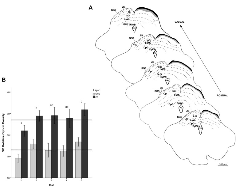
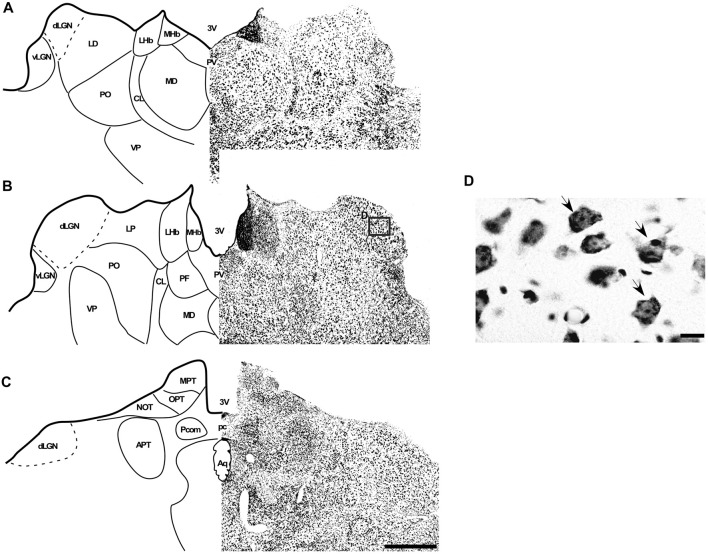
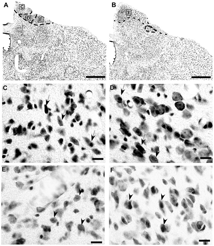
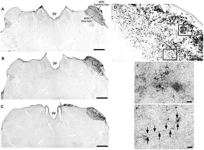
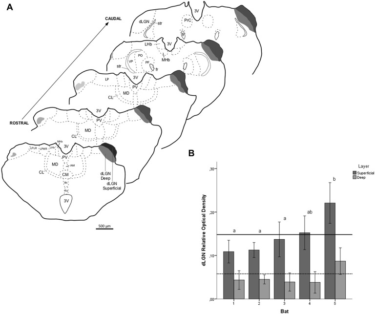
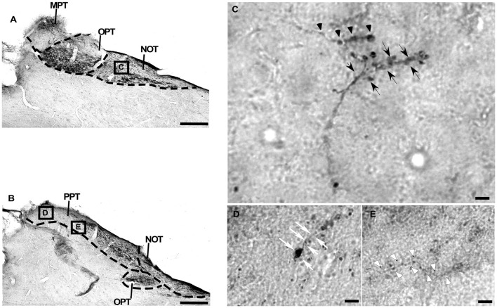
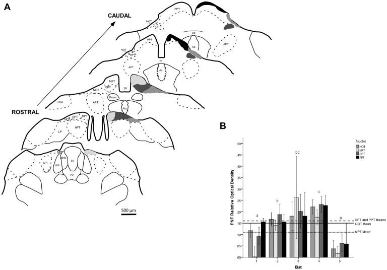
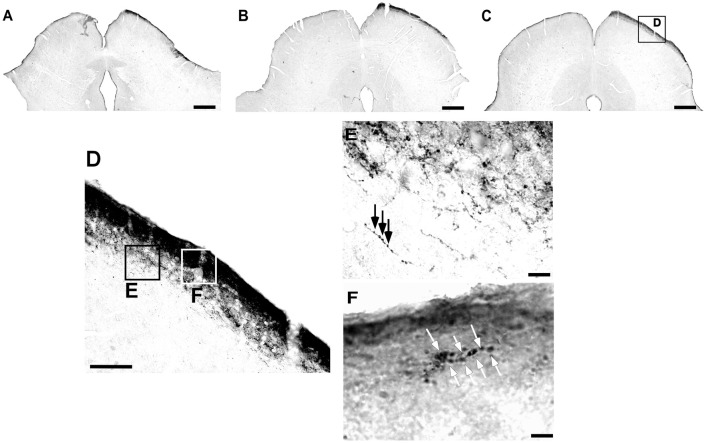
A well-developed visual system can provide significant sensory information to guide motor behavior, especially in fruit-eating bats, which usually use echolocation to navigate at high speed through cluttered environments during foraging. Relatively few studies have been performed to elucidate the organization of the visual system in bats. The present work provides an extensive morphological description of the retinal projections in the subcortical visual nuclei in the flat-faced fruit-eating bat () using anterograde transport of the eye-injected cholera toxin B subunit (CTb), followed by morphometrical and stereological analyses. Regarding the cytoarchitecture, the dorsal lateral geniculate nucleus (dLGN) was homogeneous, with no evident lamination. However, the retinal projection contained two layers that had significantly different marking intensities and a massive contralateral input. The superior colliculus (SC) was identified as a laminar structure composed of seven layers, and the retinal input was only observed on the contralateral side, targeting two most superficial layers. The medial pretectal nucleus (MPT), olivary pretectal nucleus (OPT), anterior pretectal nucleus (APT), posterior pretectal nucleus (PPT) and nucleus of the optic tract (NOT) were comprised the pretectal nuclear complex (PNT). Only the APT lacked a retinal input, which was predominantly contralateral in all other nuclei. Our results showed the morphometrical and stereological features of a bat species for the first time.
发育良好的视觉系统可以提供重要的感官信息来指导运动行为,尤其是在食果蝙蝠中,它们通常在觅食时利用回声定位在杂乱的环境中高速导航。相对较少的研究致力于阐明蝙蝠视觉系统的组织。本研究使用眼内注射霍乱毒素B亚单位(CTb)的顺行运输,随后进行形态计量学和体视学分析,对扁脸食果蝠皮层下视觉核中的视网膜投射进行了广泛的形态学描述。关于细胞结构,背外侧膝状核(dLGN)是均匀的,没有明显的分层。然而,视网膜投射包含两层,标记强度有显著差异,且有大量对侧输入。上丘(SC)被确定为一个由七层组成的分层结构,视网膜输入仅在对侧观察到,靶向最浅的两层。内侧顶盖前核(MPT)、橄榄顶盖前核(OPT)、前顶盖前核(APT)、后顶盖前核(PPT)和视束核(NOT)组成了顶盖前核复合体(PNT)。只有APT缺乏视网膜输入,在所有其他核中视网膜输入主要是对侧的。我们的结果首次展示了一种蝙蝠物种的形态计量学和体视学特征。