Medical Imaging Unit, Bioengineering Department, IRCCS Istituto di Ricerche Farmacologiche Mario Negri, Bergamo, Italy.
Department of Radiology, Ludwig-Maximilians-University Hospital Munich, Munich, Germany.
Nephrol Dial Transplant. 2018 Sep 1;33(suppl_2):ii29-ii40. doi: 10.1093/ndt/gfy163.
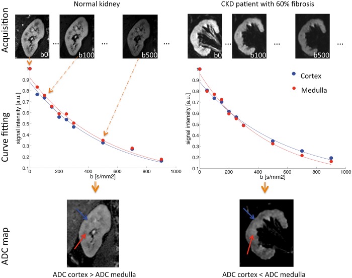
Diffusion-weighted magnetic resonance imaging (DWI) is a non-invasive method sensitive to local water motion in the tissue. As a tool to probe the microstructure, including the presence and potentially the degree of renal fibrosis, DWI has the potential to become an effective imaging biomarker. The aim of this review is to discuss the current status of renal DWI in diffuse renal diseases. DWI biomarkers can be classified in the following three main categories: (i) the apparent diffusion coefficient-an overall measure of water diffusion and microcirculation in the tissue; (ii) true diffusion, pseudodiffusion and flowing fraction-providing separate information on diffusion and perfusion or tubular flow; and (iii) fractional anisotropy-measuring the microstructural orientation. An overview of human studies applying renal DWI in diffuse pathologies is given, demonstrating not only the feasibility and intra-study reproducibility of DWI but also highlighting the need for standardization of methods, additional validation and qualification. The current and future role of renal DWI in clinical practice is reviewed, emphasizing its potential as a surrogate and monitoring biomarker for interstitial fibrosis in chronic kidney disease, as well as a surrogate biomarker for the inflammation in acute kidney diseases that may impact patient selection for renal biopsy in acute graft rejection. As part of the international COST (European Cooperation in Science and Technology) action PARENCHIMA (Magnetic Resonance Imaging Biomarkers for Chronic Kidney Disease), aimed at eliminating the barriers to the clinical use of functional renal magnetic resonance imaging, this article provides practical recommendations for future design of clinical studies and the use of renal DWI in clinical practice.
弥散加权磁共振成像(DWI)是一种对组织内局部水分子运动敏感的非侵入性方法。作为一种探测微观结构的工具,包括肾纤维化的存在和潜在程度,DWI 有可能成为一种有效的成像生物标志物。本文旨在讨论弥散性肾脏疾病中肾脏 DWI 的现状。DWI 生物标志物可分为以下三大类:(i)表观扩散系数——一种对组织中水分子扩散和微循环的综合测量;(ii)真实扩散、假性扩散和流动分数——提供扩散和灌注或管状流动的单独信息;(iii)各向异性分数——测量微观结构的方向。本文综述了在弥漫性病变中应用肾脏 DWI 的人体研究,不仅展示了 DWI 的可行性和研究内可重复性,还强调了方法标准化、额外验证和资格认证的必要性。本文还回顾了肾脏 DWI 在临床实践中的当前和未来作用,强调了其作为慢性肾脏病间质纤维化的替代和监测生物标志物的潜力,以及作为急性肾脏病炎症的替代生物标志物的潜力,这可能会影响急性移植物排斥反应中肾脏活检的患者选择。作为旨在消除功能磁共振成像在慢性肾脏病中的临床应用障碍的国际 COST(欧洲科学技术合作)行动 PARENCHIMA 的一部分,本文为未来的临床研究设计和临床实践中肾脏 DWI 的应用提供了实用建议。