Okamura Nobuyuki, Harada Ryuichi, Ishiki Aiko, Kikuchi Akio, Nakamura Tadaho, Kudo Yukitsuka
1Division of Pharmacology, Faculty of Medicine, Tohoku Medical and Pharmaceutical University, Sendai, Japan.
3Institute of Development, Aging and Cancer, Tohoku University, Sendai, Japan.
Clin Transl Imaging. 2018;6(4):305-316. doi: 10.1007/s40336-018-0290-y. Epub 2018 Jul 20.
To provide an overview on positron emission tomography (PET) imaging of tau pathology in Alzheimer's disease (AD) and other neurodegenerative disorders.
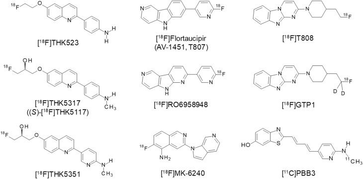
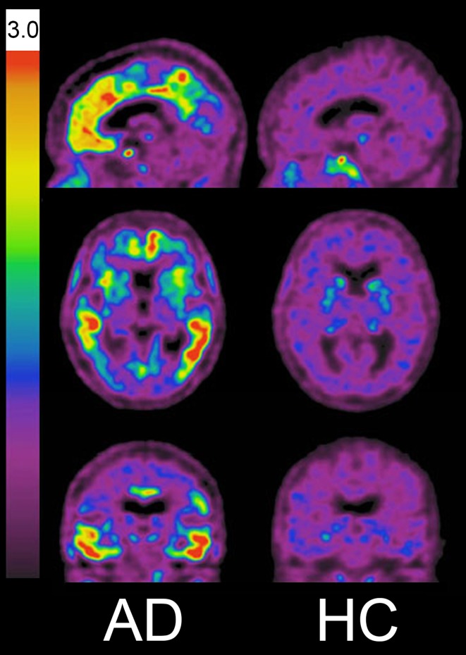
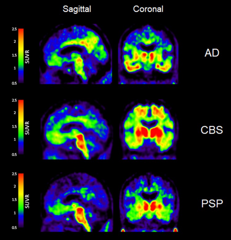
Different classes of tau tracers such as flortaucipir, THK5317, and PBB3 have been developed and utilized in previous clinical studies. In AD, the topographical distribution of tracer binding follows the known distribution of neurofibrillary tangles and is closely associated with neurodegeneration as well as the clinical phenotype of dementia. Significant retention of tracers has also been observed in the frequent site of the 4-repeat (4R) tau isoform deposits in non-AD tauopathies, such as in progressive supranuclear palsy. However, in vitro binding studies indicate that most tau tracers are less sensitive to straight tau filaments, in contrast to their high binding affinity to paired helical filaments of tau (PHF-tau). The first-generation of tau tracers shows off-target binding in the basal ganglia, midbrain, thalamus, choroid plexus, and venous sinus. Off-target binding of THK5351 to monoamine oxidase B (MAO-B) has been observed in disease-associated brain regions linked to neurodegeneration and is associated with astrogliosis in areas of misfolded protein accumulation. The second generation of tau tracers, such as [F]MK-6240, is highly selective to PHF-tau with little off-target binding and have enabled the reliable assessment of PHF-tau burden in aging and AD.
Tau PET tracers have enabled in vivo quantification of PHF-tau burden in human brains. Tau PET can help in understanding the underlying cause of dementia symptoms, and in patient selection for clinical trials of anti-dementia therapies.
综述正电子发射断层扫描(PET)在阿尔茨海默病(AD)和其他神经退行性疾病中tau病理成像的研究情况。
以往临床研究中已开发并使用了不同类型的tau示踪剂,如氟代脱氧葡萄糖、THK5317和PBB3。在AD中,示踪剂结合的拓扑分布遵循已知的神经原纤维缠结分布,与神经退行性变以及痴呆的临床表型密切相关。在非AD tau蛋白病(如进行性核上性麻痹)中,4重复(4R)tau蛋白异构体沉积的常见部位也观察到示踪剂的显著滞留。然而,体外结合研究表明,与tau蛋白配对螺旋丝(PHF-tau)的高结合亲和力相比,大多数tau示踪剂对直链tau丝的敏感性较低。第一代tau示踪剂在基底神经节、中脑、丘脑、脉络丛和静脉窦中显示出非靶向结合。在与神经退行性变相关的疾病脑区中观察到THK5351与单胺氧化酶B(MAO-B)的非靶向结合,并且在错误折叠蛋白积累区域与星形胶质细胞增生有关。第二代tau示踪剂,如[F]MK-6240,对PHF-tau具有高度选择性,几乎没有非靶向结合,能够可靠地评估衰老和AD中PHF-tau的负担。
Tau PET示踪剂能够在体内定量人脑PHF-tau的负担。Tau PET有助于理解痴呆症状的潜在原因,并为抗痴呆治疗的临床试验选择患者。