Gutiérrez Yeisson, Ott David, Töpperwien Mareike, Salditt Tim, Scherber Christoph
Institute of Landscape Ecology University of Münster Münster Germany.
Institute for X-Ray Physics University of Göttingen Göttingen Germany.
Ecol Evol. 2018 Jul 6;8(15):7717-7732. doi: 10.1002/ece3.4149. eCollection 2018 Aug.
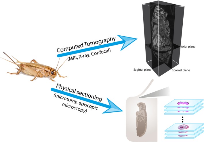
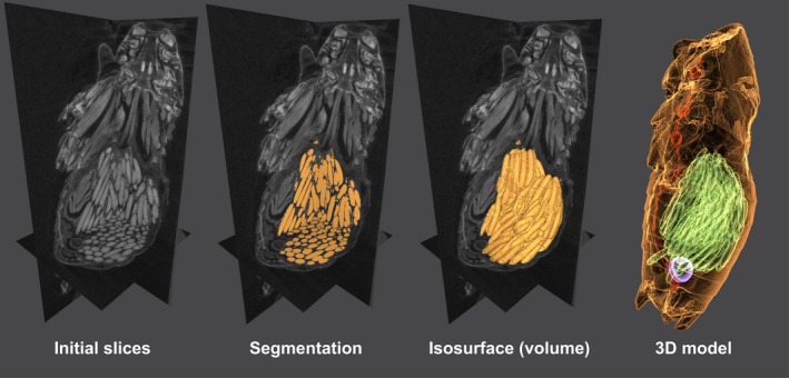
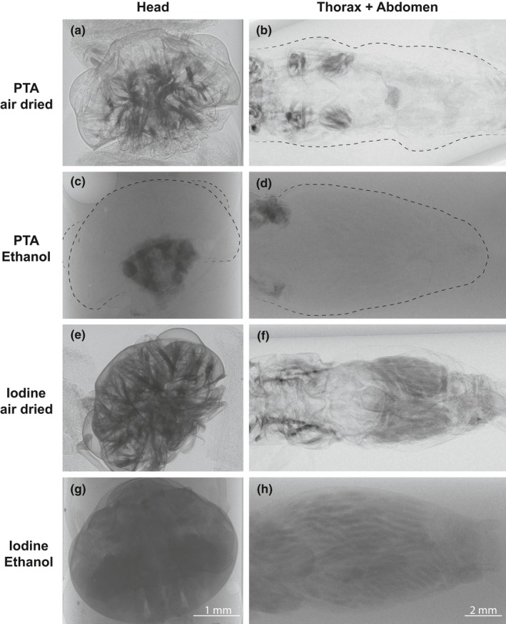
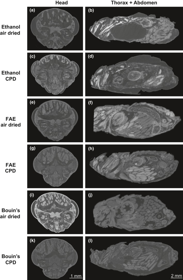
Imaging techniques are a cornerstone of contemporary biology. Over the last decades, advances in microscale imaging techniques have allowed fascinating new insights into cell and tissue morphology and internal anatomy of organisms across kingdoms. However, most studies so far provided snapshots of given reference taxa, describing organs and tissues under "idealized" conditions. Surprisingly, there is an almost complete lack of studies investigating how an organism's internal morphology changes in response to environmental drivers. Consequently, ecology as a scientific discipline has so far almost neglected the possibilities arising from modern microscale imaging techniques. Here, we provide an overview of recent developments of X-ray computed tomography as an affordable, simple method of high spatial resolution, allowing insights into three-dimensional anatomy both and . We review ecological studies using this technique to investigate the three-dimensional internal structure of organisms. In addition, we provide practical comparisons between different preparation techniques for maximum contrast and tissue differentiation. In particular, we consider the novel modality of phase contrast by self-interference of the X-ray wave behind an object (i.e., phase contrast by free space propagation). Using the cricket (L.) as model organism, we found that the combination of FAE fixative and iodine staining provided the best results across different tissues. The drying technique also affected contrast and prevented artifacts in specific cases. Overall, we found that for the interests of ecological studies, X-ray computed tomography is useful when the tissue or structure of interest has sufficient contrast that allows for an automatic or semiautomatic segmentation. In particular, we show that reconstruction schemes which exploit phase contrast can yield enhanced image quality. Combined with suitable specimen preparation and automated analysis, X-ray CT can therefore become a promising quantitative 3D imaging technique to study organisms' responses to environmental drivers, in both ecology and evolution.
成像技术是当代生物学的基石。在过去几十年中,微观成像技术的进步使人们能够对不同生物界的细胞和组织形态以及生物体的内部解剖结构有了迷人的新见解。然而,迄今为止,大多数研究提供的是特定参考分类群的快照,描述的是“理想化”条件下的器官和组织。令人惊讶的是,几乎完全缺乏研究来探究生物体的内部形态如何响应环境驱动因素而发生变化。因此,作为一门科学学科,生态学迄今为止几乎忽略了现代微观成像技术带来的可能性。在这里,我们概述了X射线计算机断层扫描技术的最新发展,它是一种经济实惠、简单的高空间分辨率方法,能够洞察生物体的三维解剖结构。我们回顾了使用该技术研究生物体三维内部结构的生态学研究。此外,我们还对不同的制备技术进行了实际比较,以实现最大对比度和组织分化。特别是,我们考虑了物体后面X射线波的自干涉产生的相衬新模态(即自由空间传播相衬)。以蟋蟀(L.)作为模式生物,我们发现FAE固定剂和碘染色的组合在不同组织中效果最佳。干燥技术也会影响对比度,并在特定情况下防止伪影。总体而言,我们发现,出于生态学研究的目的,当感兴趣的组织或结构具有足够的对比度以允许自动或半自动分割时,X射线计算机断层扫描是有用的。特别是,我们表明利用相衬的重建方案可以提高图像质量。因此,结合合适的标本制备和自动分析,X射线CT可以成为一种有前途的定量三维成像技术,用于研究生态学和进化中生物体对环境驱动因素的响应。