Zhang Ze, Wang Hui-Jing, Wang Dian-Ru, Qu Wei-Min, Huang Zhi-Li
Institutes of Brain Science and Collaborative Innovation Center for Brain Science, Department of Pharmacology, School of Basic Medical Sciences, State Key Laboratory of Medical Neurobiology and Shanghai Key Laboratory of Clinical Geriatric Medicine, Shanghai Medical College, Fudan University, Shanghai 200032, China.
Light Sci Appl. 2017 May 5;6(5):e16231. doi: 10.1038/lsa.2016.231. eCollection 2017 May.
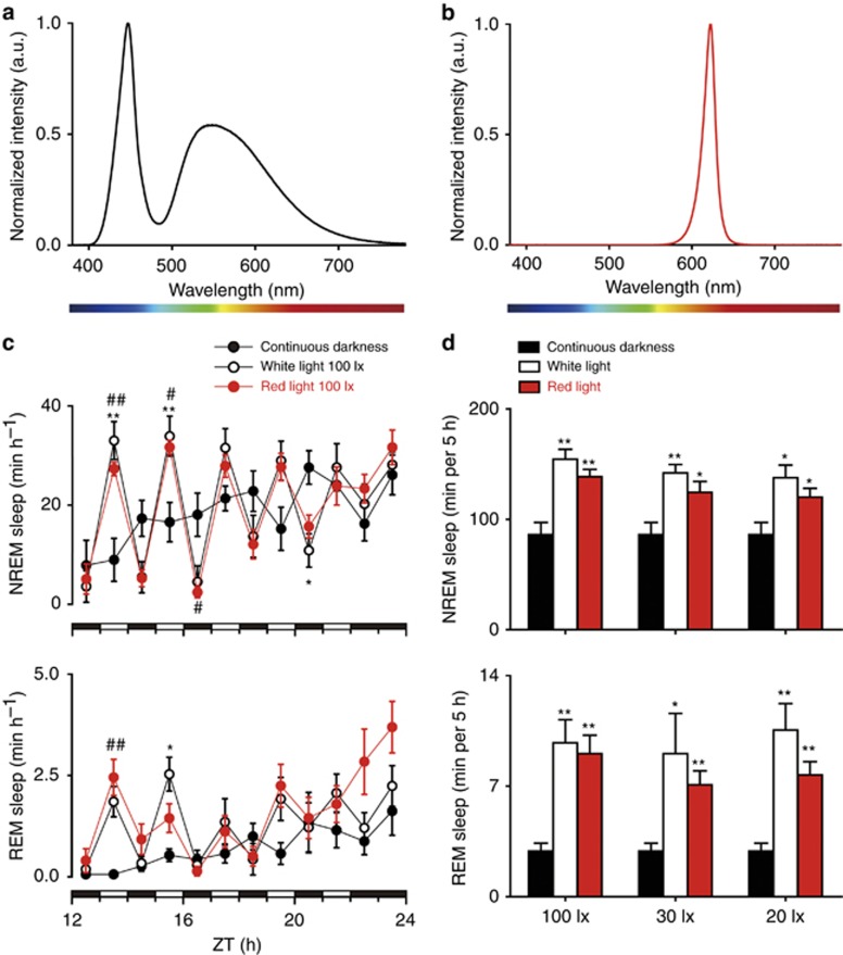
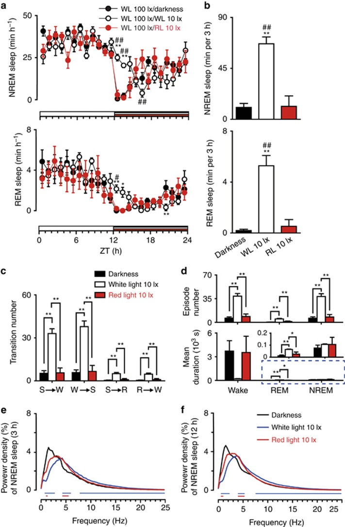
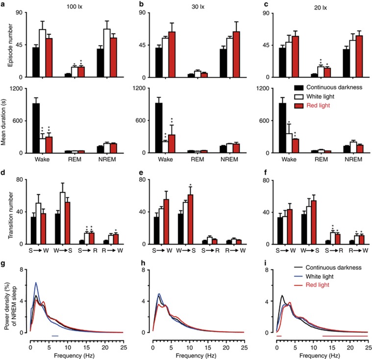
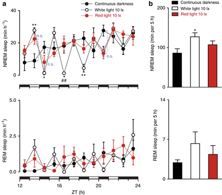
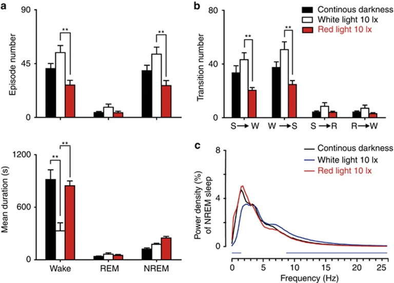
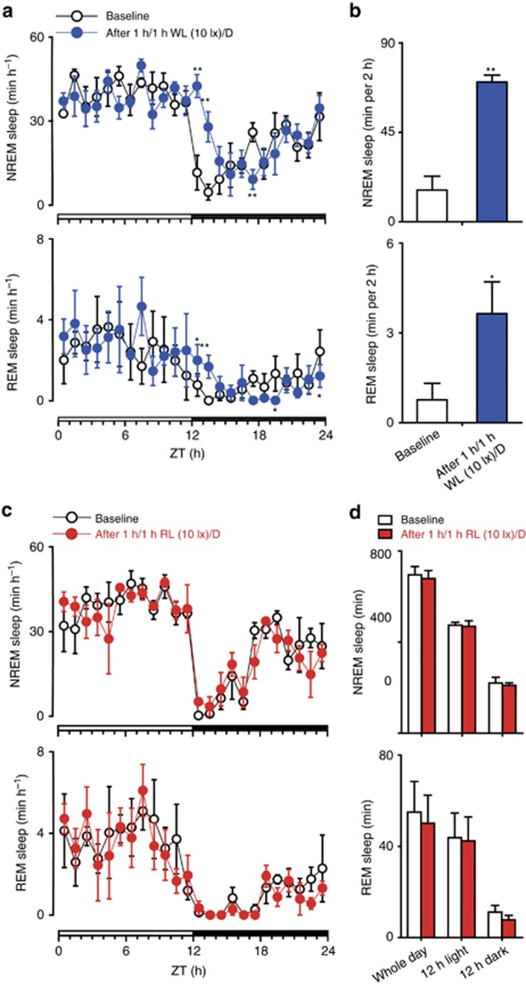
Sleep is regulated by two mechanisms: the homeostatic process and the circadian clock. Light affects sleep and alertness by entraining the circadian clock, and acutely inducing sleep/alertness, in a manner mediated by intrinsically photosensitive retinal ganglion cells. Because intrinsically photosensitive retinal ganglion cells are believed to be minimally sensitive to red light, which is widely used for illumination to reduce the photic disturbance to nocturnal animals during the dark phase. However, the appropriate intensity of the red light is unknown. In the present study, we recorded electroencephalograms and electromyograms of freely moving mice to investigate the effects of red light emitted by light-emitting diodes at different intensities and for different durations on the sleep-wake behavior of mice. White light was used as a control. Unexpectedly, red light exerted potent sleep-inducing effects and changed the sleep architecture in terms of the duration and number of sleep episodes, the stage transition, and the EEG power density when the intensity was >20 lx. Subsequently, we lowered the light intensity and demonstrated that red light at or below 10 lx did not affect sleep-wake behavior. White light markedly induced sleep and disrupted sleep architecture even at an intensity as low as 10 lx. Our findings highlight the importance of limiting the intensity of red light (⩽10 lx) to avoid optical influence in nocturnal behavioral experiments, particularly in the field of sleep and circadian research.
稳态过程和生物钟。光线通过调节生物钟以及以一种由内在光敏性视网膜神经节细胞介导的方式急性诱导睡眠/警觉性,从而影响睡眠和警觉性。由于内在光敏性视网膜神经节细胞被认为对红光的敏感性最低,而红光被广泛用于照明,以减少黑暗阶段对夜行性动物的光干扰。然而,红光的合适强度尚不清楚。在本研究中,我们记录了自由活动小鼠的脑电图和肌电图,以研究不同强度和持续时间的发光二极管发出的红光对小鼠睡眠-觉醒行为的影响。白光用作对照。出乎意料的是,当强度>20勒克斯时,红光具有强大的诱导睡眠作用,并在睡眠时长和睡眠发作次数、阶段转换以及脑电图功率密度方面改变了睡眠结构。随后,我们降低了光强度,并证明10勒克斯及以下的红光不会影响睡眠-觉醒行为。即使在低至10勒克斯的强度下,白光也会显著诱导睡眠并扰乱睡眠结构。我们的研究结果强调了在夜间行为实验中,特别是在睡眠和生物钟研究领域,限制红光强度(⩽10勒克斯)以避免光学影响的重要性。