Department of Radiology, Seoul National University Hospital, Seoul National University College of Medicine, Seoul 03080, Korea.
Department of Radiology, Samsung Medical Center, Seoul 06351, Korea.
Korean J Radiol. 2018 Sep-Oct;19(5):859-865. doi: 10.3348/kjr.2018.19.5.859. Epub 2018 Aug 6.
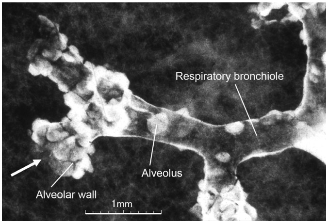
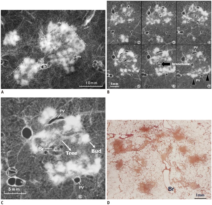
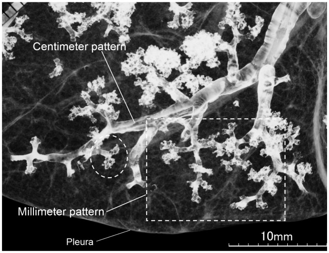
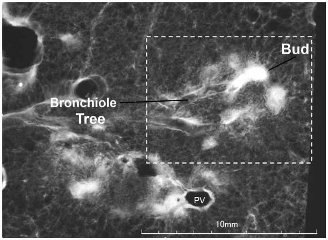
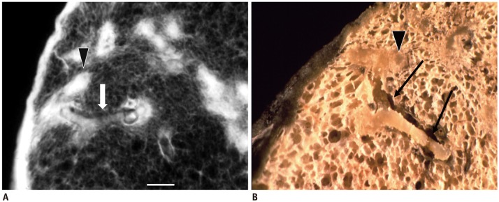
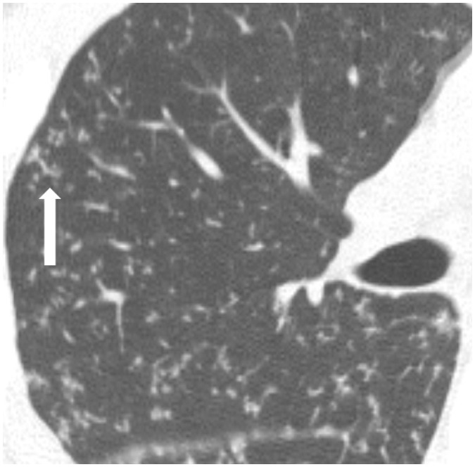
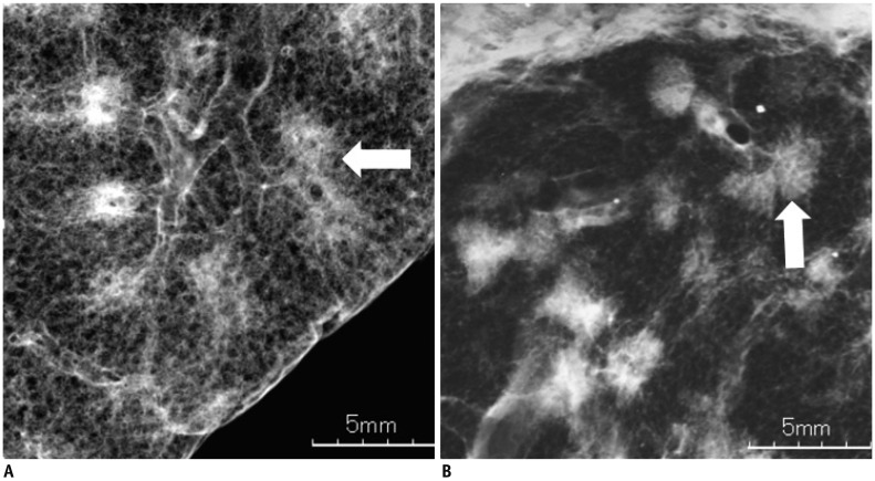
The "tree-in-bud-pattern" of images on thin-section lung CT is defined by centrilobular branching structures that resemble a budding tree. We investigated the pathological basis of the tree-in-bud lesion by reviewing the pathological specimens of bronchograms of normal lungs and contract radiographs of the post-mortem lungs manifesting active pulmonary tuberculosis. The tree portion corresponds to the intralobular inflammatory bronchiole, while the bud portion represents filling of inflammatory substances within alveolar ducts, which are larger than the corresponding bronchioles. Inflammatory bronchiole per se represents the "tree" (stem) and inflammatory alveolar ducts constitute the "buds" or clubbing. "Clusters of micronodules", seen on 7-mm thick post-mortem radiographs with tuberculosis proved to be clusters of tree-in-bud lesions within the three-dimensional space of secondary pulmonary lobule based on radiological/pathological correlation. None of the post-mortem lung specimens showed findings of lung parenchymal lymphatics involvement.
肺部 CT 薄层图像上的“树芽征”由小叶中心性分支结构组成,形似发芽的树。我们通过复习正常支气管铸型的病理标本和表现为活动性肺结核的死后肺的收缩片,研究了树芽征的病理基础。树部分对应于小叶内炎症性细支气管,而芽部分代表肺泡管内炎症物质的填充,其大于相应的细支气管。单纯的炎症性细支气管本身代表“树”(干),而炎症性肺泡管构成“芽”或棒状。在尸体放射学检查中,7mm 厚的放射片中可见到微结节簇,在放射学/病理学相关性的基础上,这些微结节簇被证明是次级肺小叶三维空间内的树芽征簇。死后肺标本均未显示肺实质淋巴管受累的表现。