• 文献检索
  • 文档翻译
  • 深度研究
  • 学术资讯
  • Suppr Zotero 插件Zotero 插件
  • 邀请有礼
  • 套餐&价格
  • 历史记录
应用&插件
Suppr Zotero 插件Zotero 插件浏览器插件Mac 客户端Windows 客户端微信小程序
定价
高级版会员购买积分包购买API积分包
服务
文献检索文档翻译深度研究API 文档MCP 服务
关于我们
关于 Suppr公司介绍联系我们用户协议隐私条款
关注我们

Suppr 超能文献

核心技术专利:CN118964589B侵权必究
粤ICP备2023148730 号-1Suppr @ 2026

文献检索

告别复杂PubMed语法,用中文像聊天一样搜索,搜遍4000万医学文献。AI智能推荐,让科研检索更轻松。

立即免费搜索

文件翻译

保留排版,准确专业,支持PDF/Word/PPT等文件格式,支持 12+语言互译。

免费翻译文档

深度研究

AI帮你快速写综述,25分钟生成高质量综述,智能提取关键信息,辅助科研写作。

立即免费体验

鉴别结核病流行地区的小尺寸(2厘米或更小)、非钙化、孤立性肺结核球与实性肺腺癌

Discriminating Small-Sized (2 cm or Less), Noncalcified, Solitary Pulmonary Tuberculoma and Solid Lung Adenocarcinoma in Tuberculosis-Endemic Areas.

作者信息

Zhang Jingping, Han Tingting, Ren Jialiang, Jin Chenwang, Zhang Ming, Guo Youmin

机构信息

Department of Radiology, The First Affiliated Hospital of Xi'an Jiaotong University, 277 West Yanta Road, Xi'an 710061, China.

GE Healthcare China, Daxing District, Tongji South Road No.1, Beijing 100176, China.

出版信息

Diagnostics (Basel). 2021 May 21;11(6):930. doi: 10.3390/diagnostics11060930.

DOI:10.3390/diagnostics11060930
PMID:34064284
原文链接:https://pmc.ncbi.nlm.nih.gov/articles/PMC8224307/
Abstract

BACKGROUND

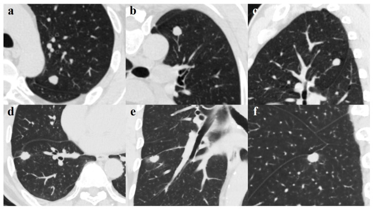
Pulmonary tuberculoma can mimic lung malignancy and thereby pose a diagnostic dilemma to clinicians. The purpose of this study was to establish an accurate, convenient, and clinically practical model for distinguishing small-sized, noncalcified, solitary pulmonary tuberculoma from solid lung adenocarcinoma.

METHODS

Thirty-one patients with noncalcified, solitary tuberculoma and 30 patients with solid adenocarcinoma were enrolled. Clinical characteristics and CT morphological features of lesions were compared between the two groups. Multivariate logistic regression analyses were applied to identify independent predictors of pulmonary tuberculoma and lung adenocarcinoma. Receiver operating characteristic (ROC) analysis was performed to investigate the discriminating efficacy.

RESULTS

The mean age of patients with tuberculoma and adenocarcinoma was 46.8 ± 12.3 years (range, 28-64) and 61.1 ± 9.9 years (range, 41-77), respectively. No significant differences were observed concerning smoking history and smoking index, underlying disease, or tumor markers between the two groups. Univariate and multivariate analyses showed age and lobulation combined with pleural indentation demonstrated excellent discrimination. The sensitivity, specificity, accuracy, and the area under the ROC curve were 87.1%, 93.3%, 90.2%, and 0.956 (95% confidence interval (CI), 0.901-1.000), respectively.

CONCLUSION

The combination of clinical characteristics and CT morphological features can be used to distinguish noncalcified, solitary tuberculoma from solid adenocarcinoma with high diagnostic performance and has a clinical application value.

摘要

背景

肺结核球可酷似肺部恶性肿瘤,从而给临床医生带来诊断难题。本研究的目的是建立一种准确、便捷且临床实用的模型,以区分小尺寸、非钙化的孤立性肺结核球与实性肺腺癌。

方法

纳入31例非钙化的孤立性结核球患者和30例实性腺癌患者。比较两组病变的临床特征和CT形态学特征。应用多因素逻辑回归分析确定肺结核球和肺腺癌的独立预测因素。进行受试者操作特征(ROC)分析以研究鉴别效能。

结果

结核球患者和腺癌患者的平均年龄分别为46.8±12.3岁(范围28 - 64岁)和61.1±9.9岁(范围41 - 77岁)。两组在吸烟史、吸烟指数、基础疾病或肿瘤标志物方面未观察到显著差异。单因素和多因素分析显示年龄以及分叶征合并胸膜凹陷具有良好的鉴别能力。ROC曲线的敏感性、特异性、准确性和曲线下面积分别为87.1%、93.3%、90.2%和0.956(95%置信区间(CI),0.901 - 1.000)。

结论

临床特征与CT形态学特征相结合可用于区分非钙化的孤立性结核球与实性腺癌,具有较高的诊断效能,具有临床应用价值。

https://cdn.ncbi.nlm.nih.gov/pmc/blobs/0a94/8224307/dc82e74f9e24/diagnostics-11-00930-g003.jpg
https://cdn.ncbi.nlm.nih.gov/pmc/blobs/0a94/8224307/821ac805da2e/diagnostics-11-00930-g001.jpg
https://cdn.ncbi.nlm.nih.gov/pmc/blobs/0a94/8224307/01de3a2be95c/diagnostics-11-00930-g002.jpg
https://cdn.ncbi.nlm.nih.gov/pmc/blobs/0a94/8224307/dc82e74f9e24/diagnostics-11-00930-g003.jpg
https://cdn.ncbi.nlm.nih.gov/pmc/blobs/0a94/8224307/821ac805da2e/diagnostics-11-00930-g001.jpg
https://cdn.ncbi.nlm.nih.gov/pmc/blobs/0a94/8224307/01de3a2be95c/diagnostics-11-00930-g002.jpg
https://cdn.ncbi.nlm.nih.gov/pmc/blobs/0a94/8224307/dc82e74f9e24/diagnostics-11-00930-g003.jpg

相似文献

1
Discriminating Small-Sized (2 cm or Less), Noncalcified, Solitary Pulmonary Tuberculoma and Solid Lung Adenocarcinoma in Tuberculosis-Endemic Areas.鉴别结核病流行地区的小尺寸(2厘米或更小)、非钙化、孤立性肺结核球与实性肺腺癌
Diagnostics (Basel). 2021 May 21;11(6):930. doi: 10.3390/diagnostics11060930.
2
Immunocyte count combined with CT features for distinguishing pulmonary tuberculoma from malignancy among non-calcified solitary pulmonary solid nodules.免疫细胞计数联合CT特征在非钙化性孤立性肺实性结节中鉴别肺结核球与恶性肿瘤
J Thorac Dis. 2023 Feb 28;15(2):386-398. doi: 10.21037/jtd-22-1024. Epub 2023 Jan 31.
3
Use of TBAg/PHA ratio in distinguishing tuberculoma from cancer in solitary pulmonary nodule or mass.结核抗原/植物血凝素比值在鉴别孤立性肺结节或肿块中的结核瘤与癌症中的应用。
Clin Respir J. 2018 Mar;12(3):1174-1181. doi: 10.1111/crj.12648. Epub 2017 May 14.
4
Radiomics nomogram for preoperative differentiation of lung tuberculoma from adenocarcinoma in solitary pulmonary solid nodule.基于影像组学的列线图模型术前鉴别肺内孤立实性结节中结核球与腺癌
Eur J Radiol. 2020 Jul;128:109022. doi: 10.1016/j.ejrad.2020.109022. Epub 2020 Apr 20.
5
[Dynamic enhanced CT evaluation of solitary pulmonary nodules].孤立性肺结节的动态增强CT评估
Zhonghua Zhong Liu Za Zhi. 2011 Apr;33(4):308-12.
6
Solitary solid pulmonary nodules: a CT-based deep learning nomogram helps differentiate tuberculosis granulomas from lung adenocarcinomas.孤立性实性肺结节:基于CT的深度学习列线图有助于鉴别结核性肉芽肿与肺腺癌。
Eur Radiol. 2020 Dec;30(12):6497-6507. doi: 10.1007/s00330-020-07024-z. Epub 2020 Jun 27.
7
Lung Adenocarcinoma at CT with 0.25-mm Section Thickness and a 2048 Matrix: High-Spatial-Resolution Imaging for Predicting Invasiveness.CT 扫描采用 0.25 毫米层厚和 2048 矩阵:高空间分辨率成像预测侵袭性。
Radiology. 2020 Nov;297(2):462-471. doi: 10.1148/radiol.2020201911. Epub 2020 Sep 8.
8
CT-based radiomics signature for differentiating solitary granulomatous nodules from solid lung adenocarcinoma.基于 CT 的放射组学特征区分孤立性肉芽肿性结节与实性肺腺癌。
Lung Cancer. 2018 Nov;125:109-114. doi: 10.1016/j.lungcan.2018.09.013. Epub 2018 Sep 17.
9
[The value of CT and MRI in differentiating malignant nodule from tuberculoma].[CT与MRI在鉴别恶性结节与结核瘤中的价值]
Zhonghua Jie He He Hu Xi Za Zhi. 1995 Aug;18(4):218-20, 255.
10
Distinguishing small primary lung cancer from pulmonary tuberculoma using 64-slices multidetector CT.使用64层螺旋CT鉴别小原发性肺癌与肺结核球
J Med Assoc Thai. 2012 Apr;95(4):574-82.

引用本文的文献

1
The diagnostic value of CT-based radiomics nomogram for solitary indeterminate smoothly marginated solid pulmonary nodules.基于CT的影像组学列线图对孤立性边界光滑的实性肺结节的诊断价值
Front Oncol. 2024 Jul 2;14:1427404. doi: 10.3389/fonc.2024.1427404. eCollection 2024.
2
EBUS-GS with the GeneXpert MTB/RIF assay for diagnosis of Mycobacterium tuberculosis infection of isolated pulmonary nodules.超声支气管镜引导下经支气管针吸活检术联合 GeneXpert MTB/RIF 检测对孤立性肺结节分枝杆菌结核感染的诊断价值。
Eur J Med Res. 2023 Sep 23;28(1):370. doi: 10.1186/s40001-023-01331-z.
3
Radiomics nomogram for preoperative differentiation of pulmonary mucinous adenocarcinoma from tuberculoma in solitary pulmonary solid nodules.

本文引用的文献

1
Solitary solid pulmonary nodules: a CT-based deep learning nomogram helps differentiate tuberculosis granulomas from lung adenocarcinomas.孤立性实性肺结节:基于CT的深度学习列线图有助于鉴别结核性肉芽肿与肺腺癌。
Eur Radiol. 2020 Dec;30(12):6497-6507. doi: 10.1007/s00330-020-07024-z. Epub 2020 Jun 27.
2
Evaluation of the diagnostic efficacy of F-Fluorine-2-Deoxy-D-Glucose PET/CT for lung cancer and pulmonary tuberculosis in a Tuberculosis-endemic Country.评估 F-氟代-2-脱氧-D-葡萄糖 PET/CT 对结核病高发国家肺癌和肺结核的诊断效能。
Cancer Med. 2020 Feb;9(3):931-942. doi: 10.1002/cam4.2770. Epub 2019 Dec 13.
3
基于影像组学的列线图模型术前鉴别肺内孤立实性结节中的黏液型肺腺癌与结核瘤。
BMC Cancer. 2023 Mar 21;23(1):261. doi: 10.1186/s12885-023-10734-4.
4
Immunocyte count combined with CT features for distinguishing pulmonary tuberculoma from malignancy among non-calcified solitary pulmonary solid nodules.免疫细胞计数联合CT特征在非钙化性孤立性肺实性结节中鉴别肺结核球与恶性肿瘤
J Thorac Dis. 2023 Feb 28;15(2):386-398. doi: 10.21037/jtd-22-1024. Epub 2023 Jan 31.
5
Identifying Solitary Granulomatous Nodules from Solid Lung Adenocarcinoma: Exploring Robust Image Features with Cross-Domain Transfer Learning.从实性肺腺癌中识别孤立性肉芽肿结节:利用跨域迁移学习探索稳健的图像特征
Cancers (Basel). 2023 Jan 31;15(3):892. doi: 10.3390/cancers15030892.
6
Differential diagnosis of pulmonary nodular mucinous adenocarcinoma and tuberculoma with dynamic CT: a retrospective study.动态CT对肺结节状黏液腺癌与结核瘤的鉴别诊断:一项回顾性研究
J Thorac Dis. 2022 Apr;14(4):1225-1231. doi: 10.21037/jtd-22-372.
Clinical Significance of Pleural Attachment and Indentation of Subsolid Nodule Lung Cancer.
实性肺小结节肺癌胸膜附着和凹陷的临床意义。
Cancer Res Treat. 2019 Oct;51(4):1540-1548. doi: 10.4143/crt.2019.057. Epub 2019 Mar 25.
4
Perinodular and Intranodular Radiomic Features on Lung CT Images Distinguish Adenocarcinomas from Granulomas.肺 CT 图像的周围和结节内放射组学特征可区分腺癌和肉芽肿
Radiology. 2019 Mar;290(3):783-792. doi: 10.1148/radiol.2018180910. Epub 2018 Dec 18.
5
Lung Adenocarcinoma: CT Features Associated with Spread through Air Spaces.肺腺癌:与空气传播途径有关的 CT 特征。
Radiology. 2018 Dec;289(3):831-840. doi: 10.1148/radiol.2018180431. Epub 2018 Sep 4.
6
Tree-in-Bud Pattern of Pulmonary Tuberculosis on Thin-Section CT: Pathological Implications.树芽征在肺结核 CT 中的表现:病理学意义。
Korean J Radiol. 2018 Sep-Oct;19(5):859-865. doi: 10.3348/kjr.2018.19.5.859. Epub 2018 Aug 6.
7
Risk of active tuberculosis among people with diabetes mellitus: systematic review and meta-analysis.糖尿病患者患活动性肺结核的风险:系统评价和荟萃分析。
Trop Med Int Health. 2018 Oct;23(10):1058-1070. doi: 10.1111/tmi.13133. Epub 2018 Aug 29.
8
Lung Cancer Screening, Version 3.2018, NCCN Clinical Practice Guidelines in Oncology.肺癌筛查,版本 3.2018,NCCN 肿瘤学临床实践指南。
J Natl Compr Canc Netw. 2018 Apr;16(4):412-441. doi: 10.6004/jnccn.2018.0020.
9
Association between diabetes mellitus and active tuberculosis: A systematic review and meta-analysis.糖尿病与活动性肺结核之间的关联:一项系统评价与荟萃分析。
PLoS One. 2017 Nov 21;12(11):e0187967. doi: 10.1371/journal.pone.0187967. eCollection 2017.
10
Evaluation of the solitary pulmonary nodule: size matters, but do not ignore the power of morphology.孤立性肺结节的评估:大小很重要,但不要忽视形态学的作用。
Insights Imaging. 2018 Feb;9(1):73-86. doi: 10.1007/s13244-017-0581-2. Epub 2017 Nov 15.